Copyright
©The Author(s) 2020.
World J Clin Cases. Dec 6, 2020; 8(23): 6009-6015
Published online Dec 6, 2020. doi: 10.12998/wjcc.v8.i23.6009
Published online Dec 6, 2020. doi: 10.12998/wjcc.v8.i23.6009
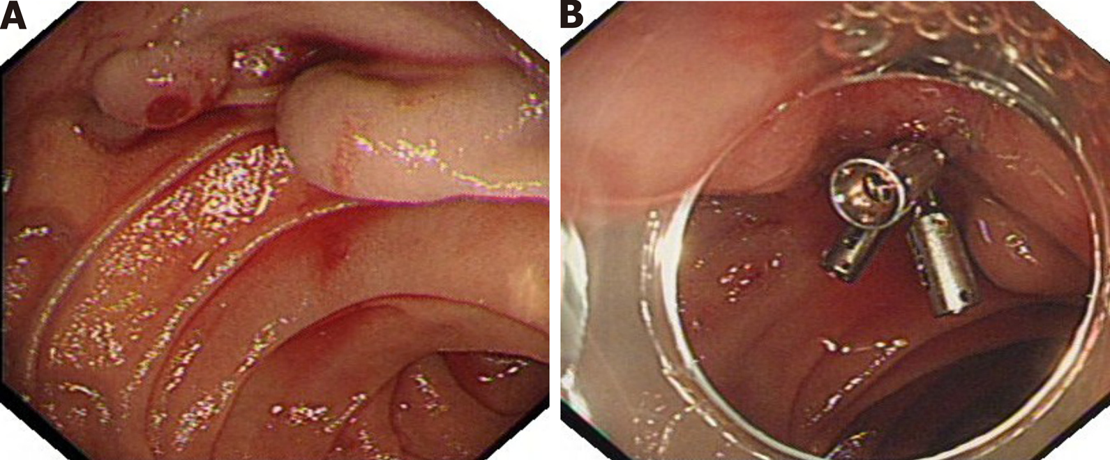
Figure 1 The first endoscopic examination found that the patient had duodenal variceal bleeding.
A: A plurality of tortuous gray-blue varicose veins are seen in the horizontal part of duodenum, with the thickest diameter of about 0.7 cm, active bleeding can be seen on the surface of varicose veins; B: The surface of varicose veins is clamped by metal clips, and bleeding stops.
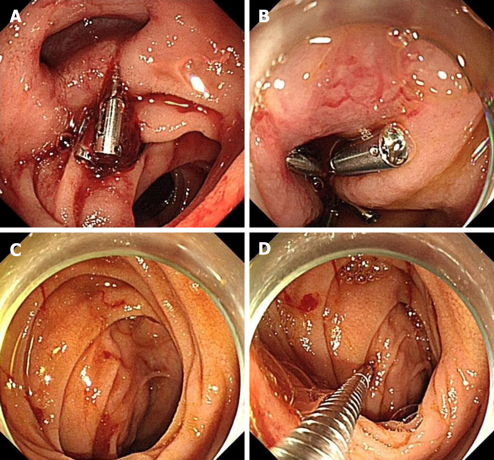
Figure 2 Endoscopic examination and hemostatic treatment of the patient in our hospital.
A: The horizontal part of duodenum can see retention of metal clip and local active bleeding; B: The bleeding position can be closed by metal clip again. Blood vessels meandering with it can be seen around it; C: Active bleeding stops. Hemispherical submucosal protuberance can be seen around bleeding; D: The boundary is unclear. The protuberance contacted by biopsy forceps is hard.
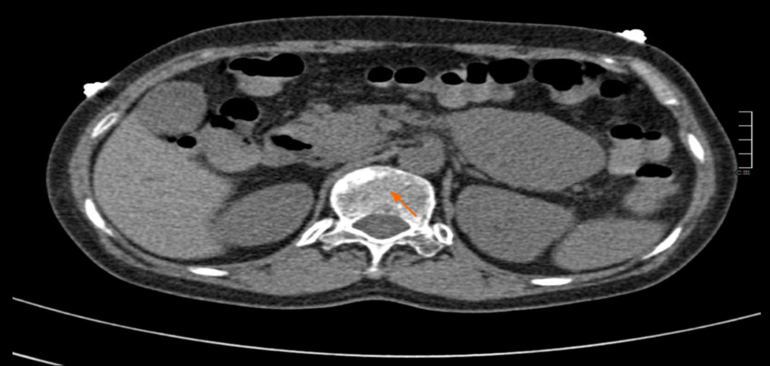
Figure 3 Abdominal computed tomography: mass soft tissue density shadow in the horizontal part of duodenum.
With a size of about 7.0 cm × 4.8 cm × 5.7 cm (orange arrow).
Figure 4 The resected mass and its pathological morphology under microscope.
A: Gross specimen; B: Microscopic tumor morphology (hematoxylin and eosin 40 ×); C: Several tortuous dilated blood vessels were found in submucosa, some of which were congested, which conformed to the pathological manifestations of varicose veins, and some of which were occluded by compression.
Figure 5 Immunohistochemistry showed CD34+ and CD117+, discovered on gastrointestinal stromal tumor-1.
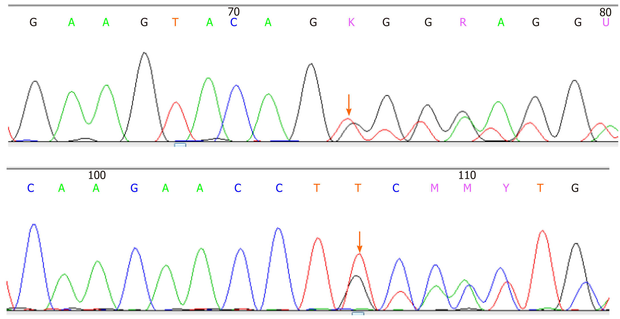
Figure 6 C-KIT gene exon-11 sequencing results: c.
1669_1674delTGGAAG(p.W557_K558del) mutation.
- Citation: Li DH, Liu XY, Xu LB. Duodenal giant stromal tumor combined with ectopic varicose hemorrhage: A case report. World J Clin Cases 2020; 8(23): 6009-6015
- URL: https://www.wjgnet.com/2307-8960/full/v8/i23/6009.htm
- DOI: https://dx.doi.org/10.12998/wjcc.v8.i23.6009


