Copyright
©The Author(s) 2020.
World J Clin Cases. Dec 6, 2020; 8(23): 5988-5998
Published online Dec 6, 2020. doi: 10.12998/wjcc.v8.i23.5988
Published online Dec 6, 2020. doi: 10.12998/wjcc.v8.i23.5988
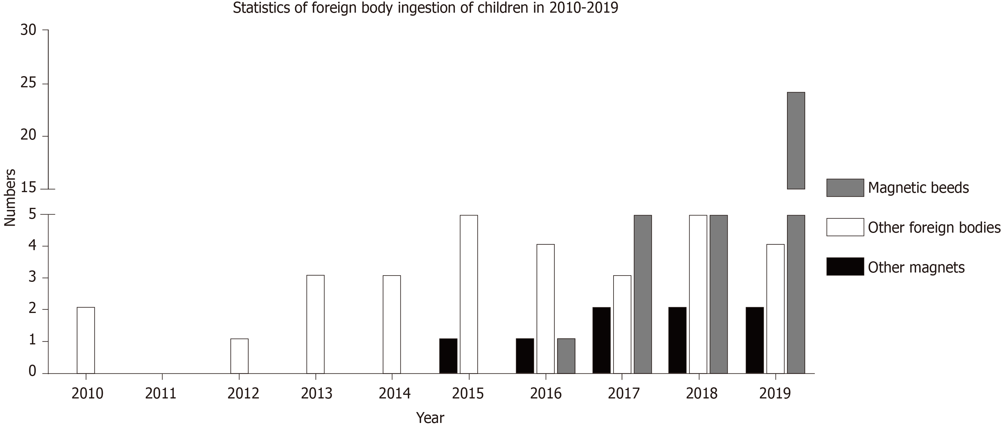
Figure 1 Statistics of ingested foreign body cases of children in our department from 2010 to 2019 (data from January to April 2020 are not included).
Figure 2 Magnetic beads adsorbed between the esophagus and trachea, causing an esophagotracheal fistula.
A: Two shadows from the magnetic beads can be seen on the chest radiograph; B: One magnetic bead at the opening of the left bronchus of the tracheal carina can be seen under the tracheoscope; C: After two magnetic beads were removed through the trachea, gastroscopy was performed to assess the esophagus, and a fistula (black arrow) was visible.
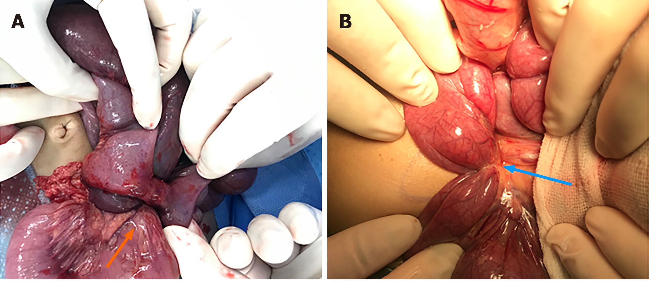
Figure 3 Intraoperative photos of two children with shock.
A: Several disc magnets adsorbed together, forming a column fistula between two small intestines (orange arrow shows) and resulting in ischemic necrosis of the compressed intestine; B: Inflammatory tissue hyperplasia near the fistula, which formed a strap (blue arrow shows) that compressed the proximal small intestine.
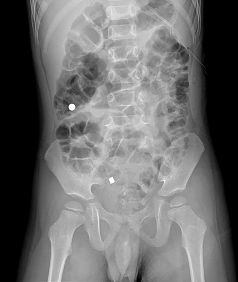
Figure 4 One patient discharged the ingested magnets at different times after ingesting the magnets at different times.
The patient was 4 years old. He swallowed two disk magnets at different times, without gastrointestinal symptoms. One magnet was excreted 3 d after swallowing, while the other was excreted 6 d after swallowing.
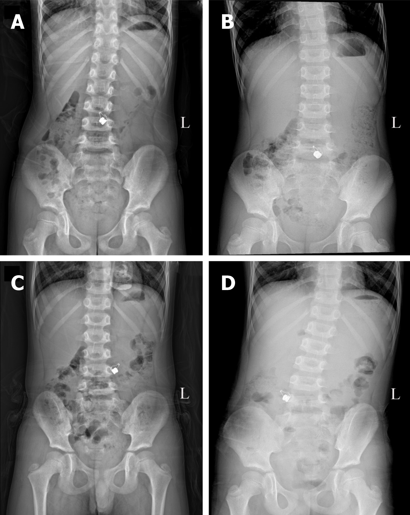
Figure 5 A 6-year-old patient swallowed two disk magnets by mistake, and presented without gastrointestinal symptoms.
The abdominal standing radiograph was obtained during conservative treatment for 4 d (The above Figure A-D showed the abdominal standing radiograph from day 1 to day 4). A magnetic shadow located in the middle and lower abdomen was observed, with obvious relocation between the images. However, two magnets were found in the stomach during operation.
- Citation: Cai DT, Shu Q, Zhang SH, Liu J, Gao ZG. Surgical treatment of multiple magnet ingestion in children: A single-center study. World J Clin Cases 2020; 8(23): 5988-5998
- URL: https://www.wjgnet.com/2307-8960/full/v8/i23/5988.htm
- DOI: https://dx.doi.org/10.12998/wjcc.v8.i23.5988


