Copyright
©The Author(s) 2020.
World J Clin Cases. Dec 6, 2020; 8(23): 5902-5917
Published online Dec 6, 2020. doi: 10.12998/wjcc.v8.i23.5902
Published online Dec 6, 2020. doi: 10.12998/wjcc.v8.i23.5902
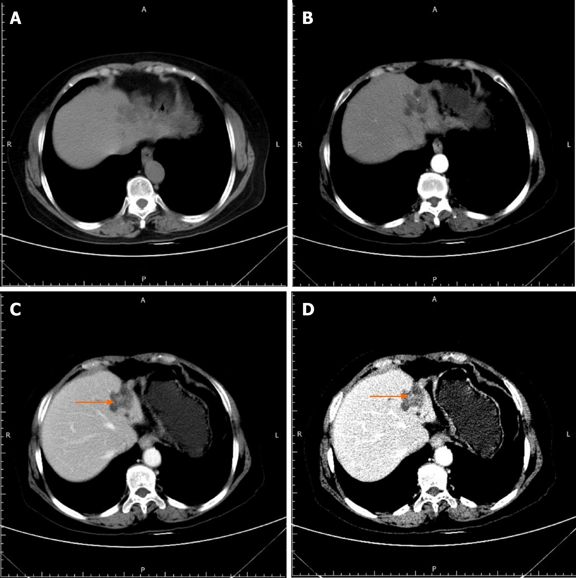
Figure 1 Computed tomography images of patient No.
1 who was diagnosed with intratubular growth-type intrahepatic cholangiocarcinoma. The orange arrow indicates a 0.8 cm faintly enhanced nodular intrahepatic cholangiocarcinoma lesion located in the dilated intrahepatic bile duct (B2/B3) of the atrophic left lobe in the portal (C) and delayed (D) phases of multidetector-row computed tomography.
Figure 2 Computed tomography images of patient No.
2 who was diagnosed with hepatocellular carcinoma with bile duct tumor thrombus. The orange arrow indicates multiple spot-like enhanced hepatocellular carcinoma lesions located within dilated B2/B3 (A), and a metastatic lesion was also detected in the right lung (B).
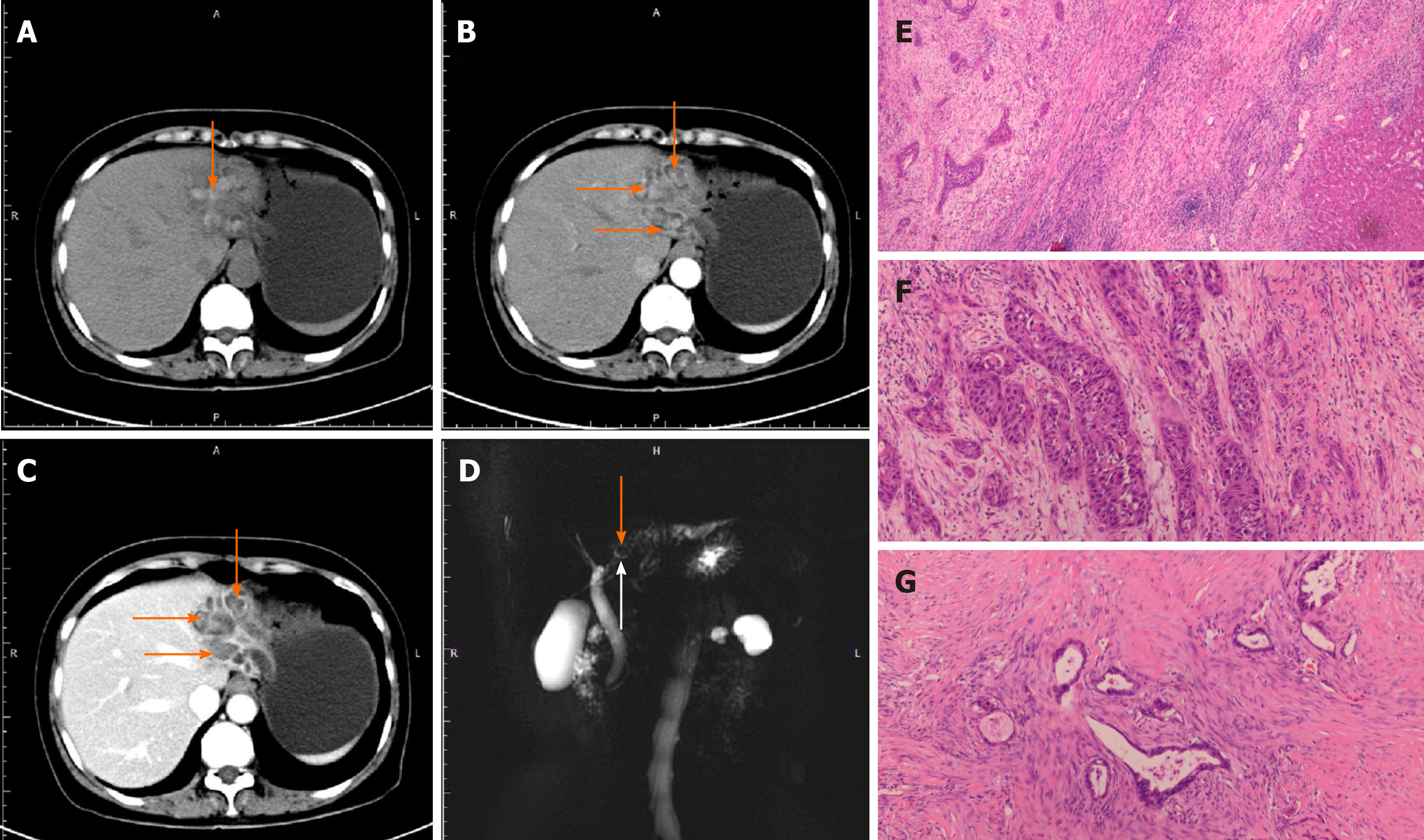
Figure 3 Computed tomography (A-C) and magnetic resonance cholangiopancreatography (D) images of patient No.
3 with adenosquamous carcinoma of the liver. A-C: The orange arrow indicates stones in the left intrahepatic bile ducts; D: The white arrow indicates that the FIHS was located at the confluence of B2/B3/B4; E-G: Pathological manifestations of adenosquamous carcinoma (ASC). Microscopically, ASC consisted of two components: adenocarcinoma (F) and squamous cell carcinoma (G).
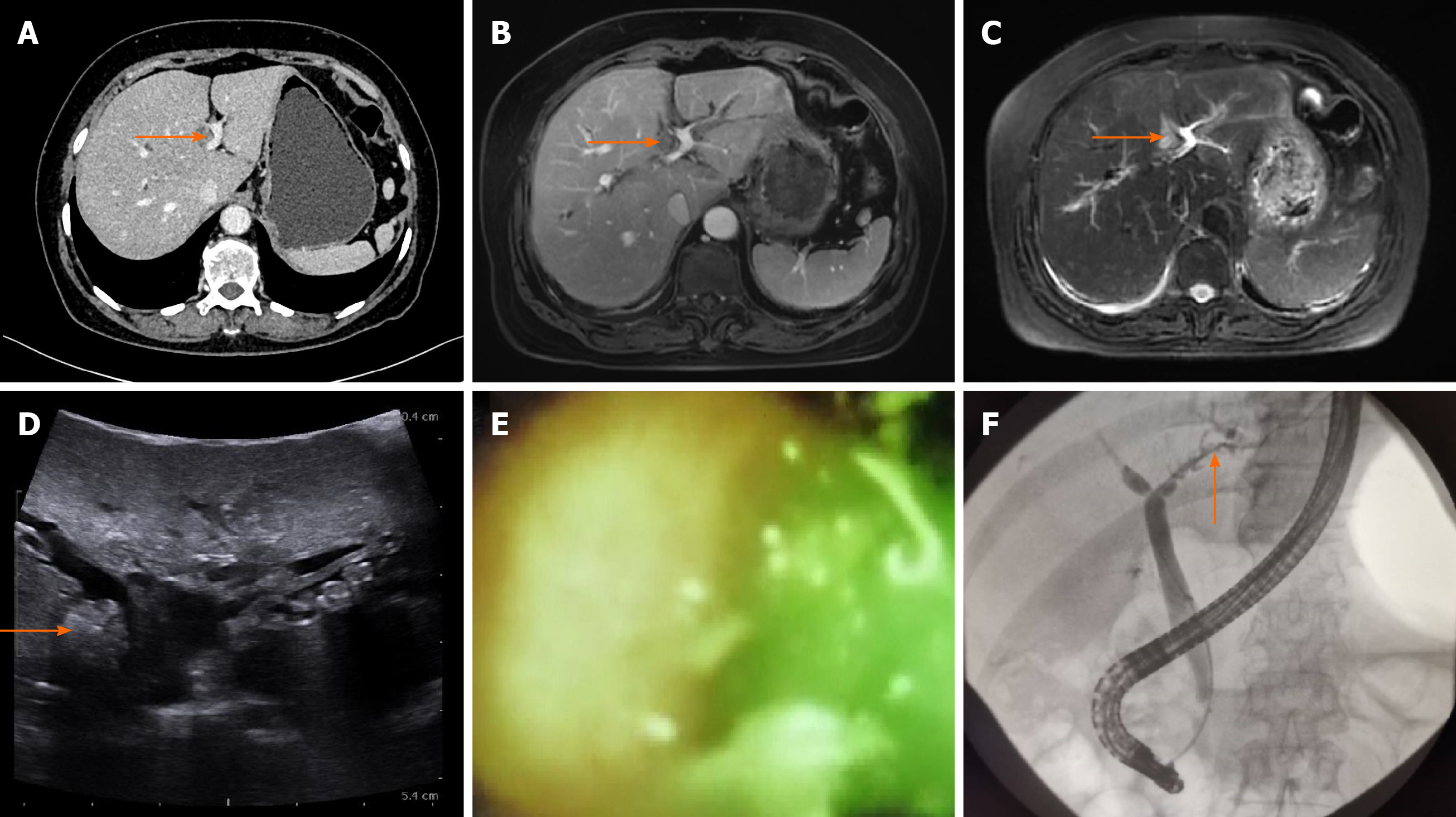
Figure 4 Multidetector-row computed tomography, magnetic resonance imaging, ultrasound, and endoscopic retrograde cholangiopancreatography images of patient No.
4. A: The orange arrow in the computed tomography scan image shows dilated B4; B and C: T1- (B) and T2-weighted (C) magnetic resonance imaging scans show low and high signals in B4, respectively; D: The ultrasound shows a 1.0 cm × 1.0 cm mass-like high-echo lesion in B4 (orange arrow); E and F: Endoscopic retrograde cholangiopancreatography with SpyGlass failed to enter B4 but detected small stones and batt-like exudate in the peripheral part of the left hepatic duct.
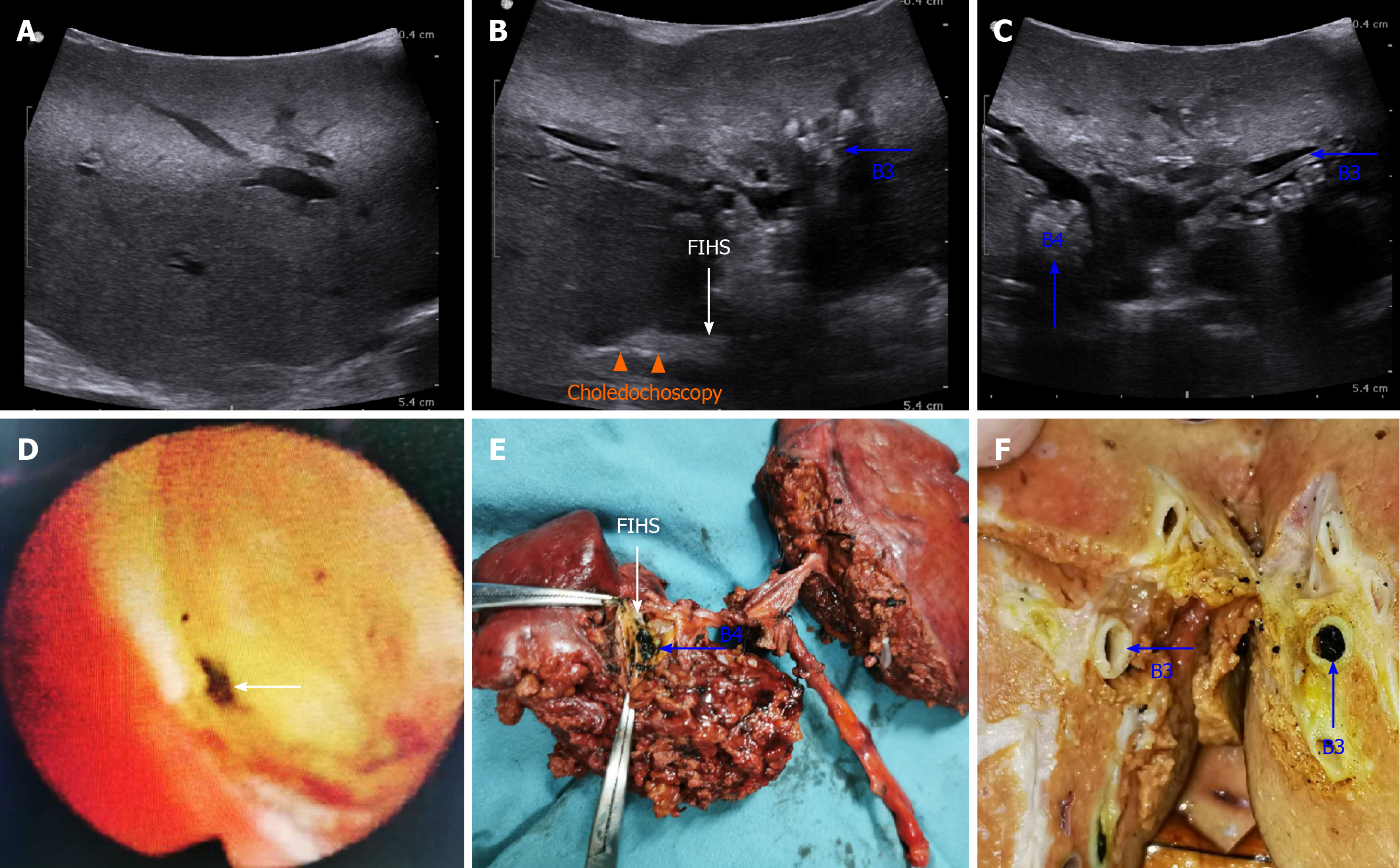
Figure 5 Intraoperative ultrasonography images of patient No.
4 (A-C). A: No dilation or stones could be detected in the intrahepatic bile ducts of the right lobe; B: Combined applications of intraoperative ultrasonography and choledochoscopy confirmed that the focal intrahepatic strictures (FIHS) was located between the peripheral part of the left hepatic duct (LHD) and the confluence of B2/B3/B4 (orange triangles). Blue arrows indicate intrahepatic stones in B3 and B4 (B and C); D: Choledochoscopy indicated FIHS at the peripheral part of the LHD; E: Specimen of the left lobe. FIHS was located at the confluence of B2/B3/B4, and stones could be seen in B4 (E) and B3 (F).
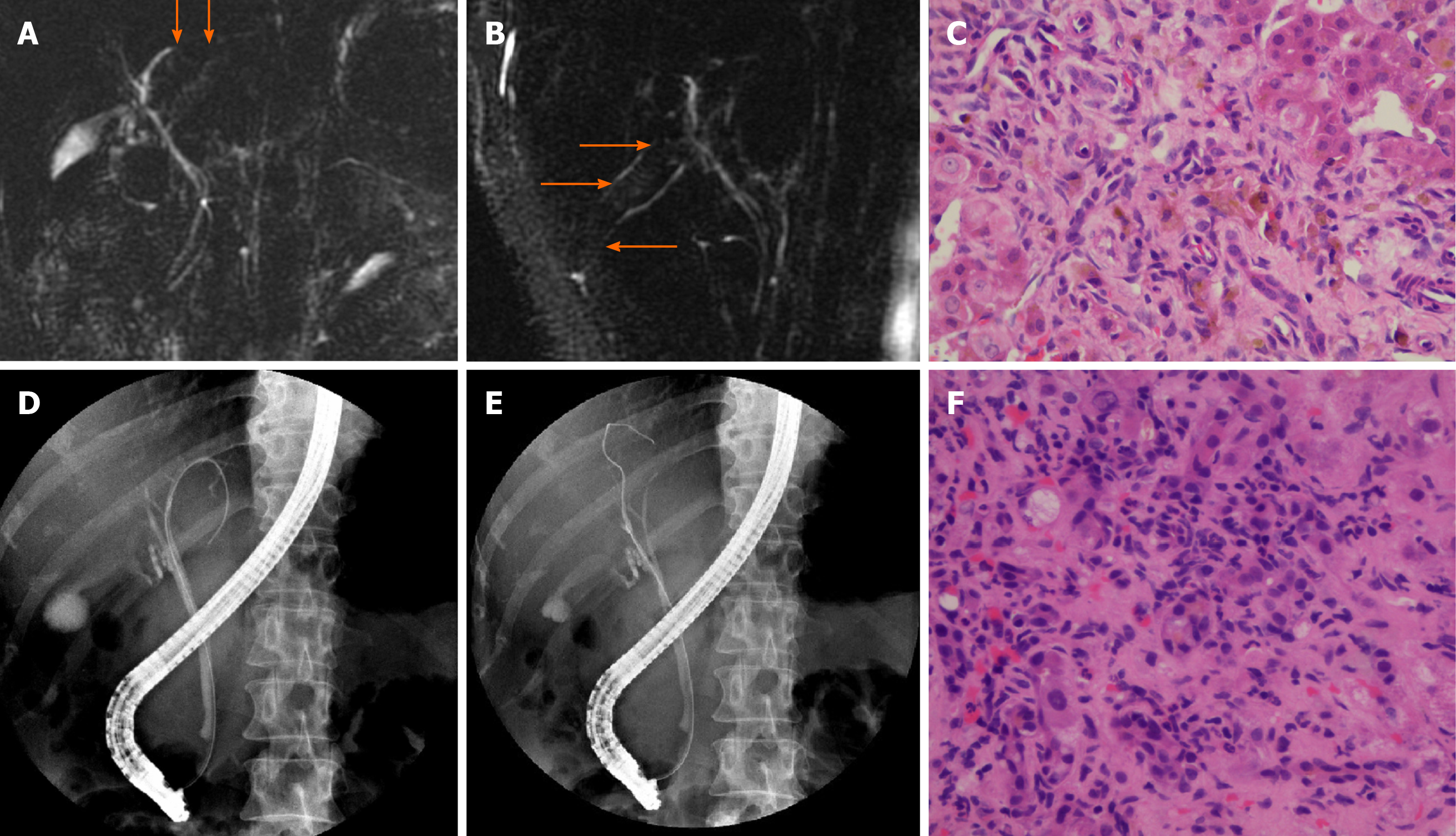
Figure 6 Magnetic resonance cholangiopancreatography images (A and B) and pathological manifestations (C) of patient No.
5 with small duct primary sclerosing cholangitis. Magnetic resonance cholangiopancreatography showed suspected multiple strictures within the biliary tree (orange arrow). Pathological examination showed preserved lobular architecture, moderate fibrosis and inflammatory processes with proliferation of ductules and feathery degeneration of hepatocytes (C). Endoscopic retrograde cholangiopancreatography (ERCP) images (D and E) and pathological manifestations (F) of patient No. 6# with AIH. ERCP showed multiple strictures of intrahepatic bile ducts in both lobes (D and E). Pathological examination showed typical chronic cholestatic hepatitis and infiltration of lymphocytes, mononuclear cells and plasma cells in the periportal area (F).
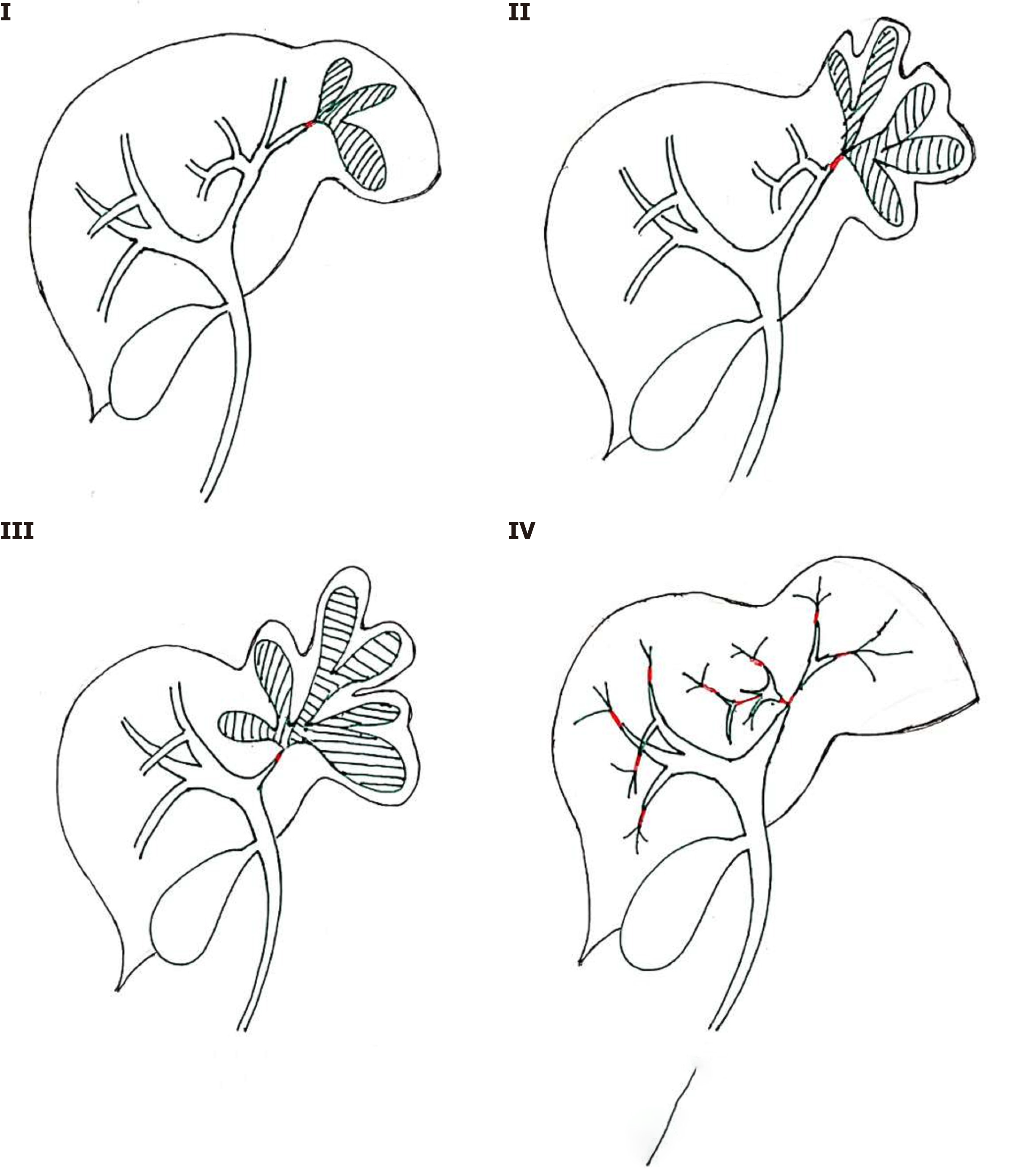
Figure 7 Our proposed focal intrahepatic strictures classification system.
Type I: Focal intrahepatic strictures (FIHS) located within one segment of the liver (A); Type II: FIHS located at the confluence of the bile ducts of one segment or two adjacent segments (B); Type III: FIHS connected to the left or right hepatic duct (C); and Type IV: Multiple FIHS located in both lobes of the liver (D).
- Citation: Zhou D, Zhang B, Zhang XY, Guan WB, Wang JD, Ma F. Focal intrahepatic strictures: A proposal classification based on diagnosis-treatment experience and systemic review. World J Clin Cases 2020; 8(23): 5902-5917
- URL: https://www.wjgnet.com/2307-8960/full/v8/i23/5902.htm
- DOI: https://dx.doi.org/10.12998/wjcc.v8.i23.5902


