Copyright
©The Author(s) 2020.
World J Clin Cases. Nov 26, 2020; 8(22): 5809-5815
Published online Nov 26, 2020. doi: 10.12998/wjcc.v8.i22.5809
Published online Nov 26, 2020. doi: 10.12998/wjcc.v8.i22.5809
Figure 1 Stomach computed tomography images.
A: Plain scan; B: Arterial phase; C: Venous phase.
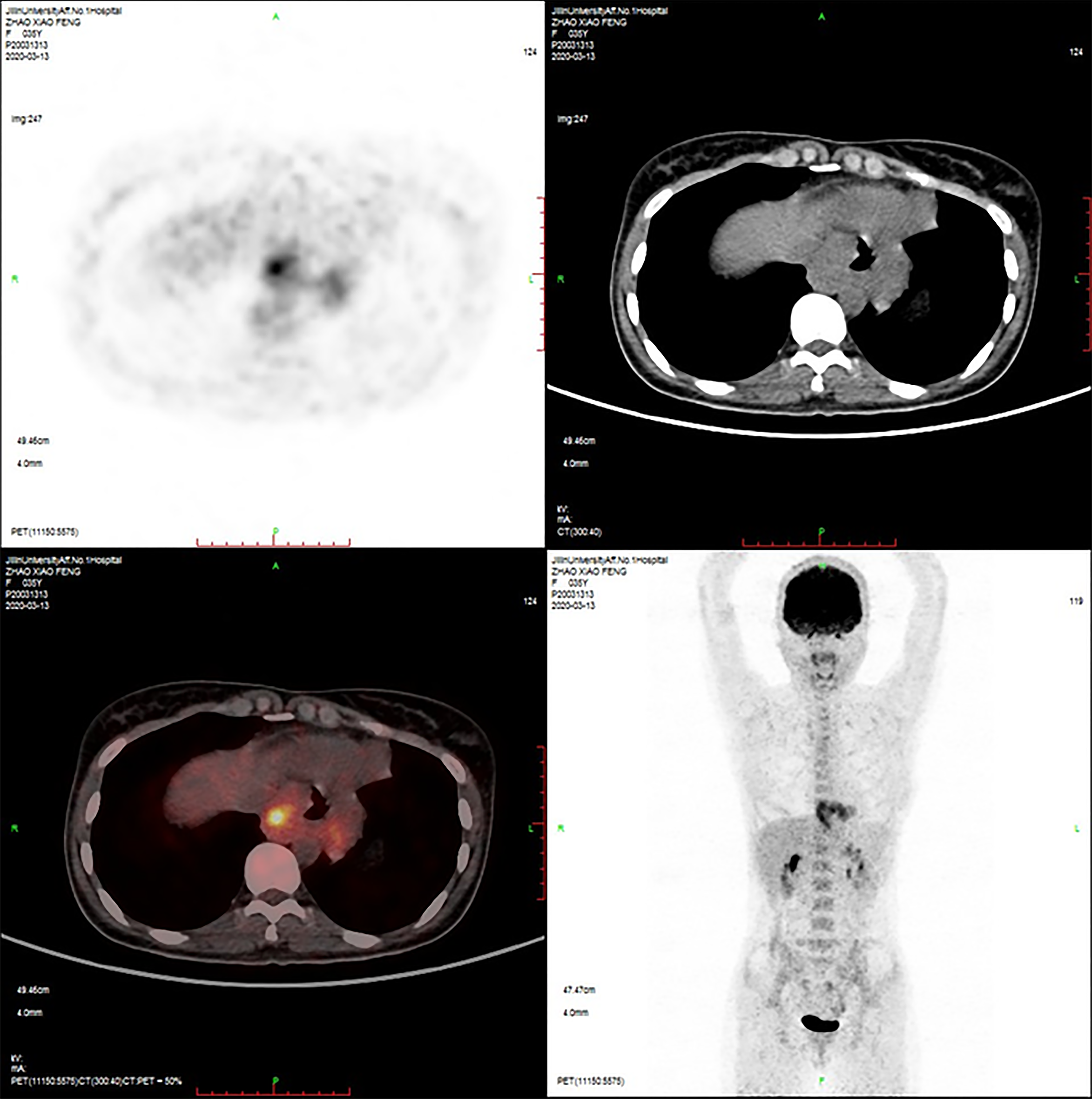
Figure 2 Positron emission tomography–computed tomography image.
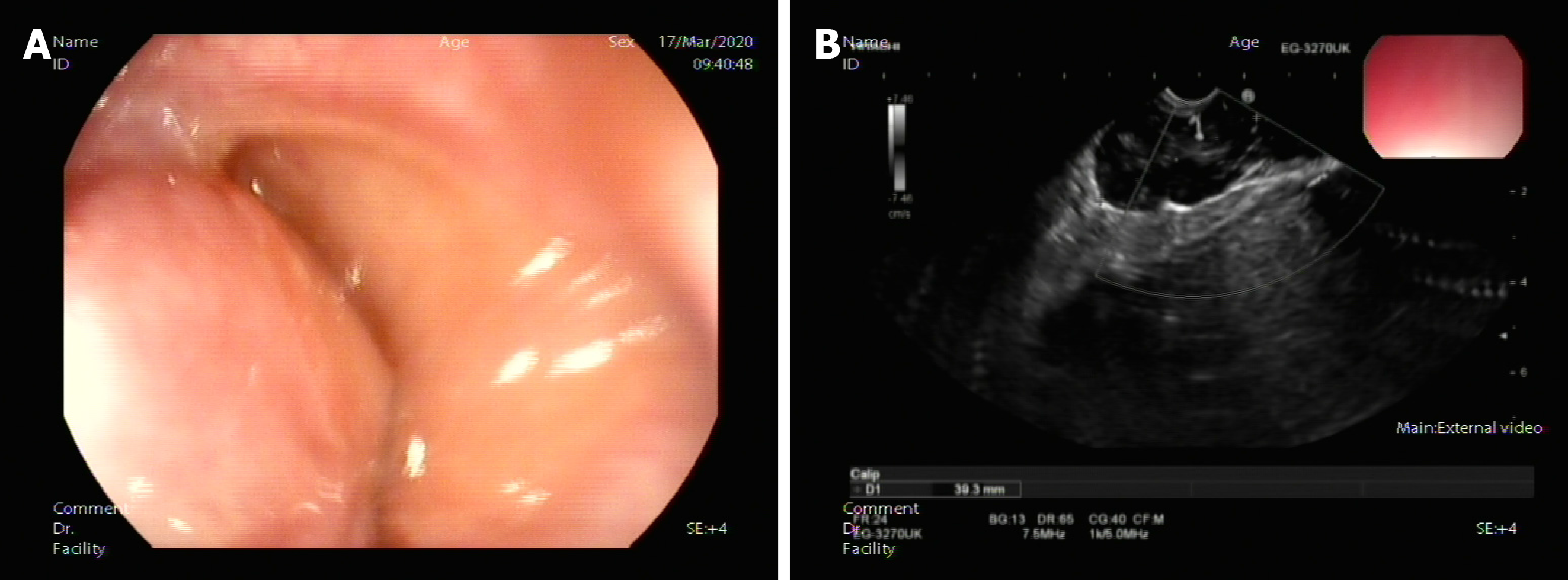
Figure 3 Endoscopic ultrasound examined the lower part of the esophagus near the cardia, and a hypoechoic mass was seen close to the esophageal wall growing outward.
A: Large uplift of the cardia under endoscopy; B: Endoscopic ultrasonography image.
Figure 4 Pathological examination.
A: Tissue specimen aspirated by endoscopic ultrasonography–fine needle aspiration (FNA); B: Cytological examination of FNA biopsy of tumor (hematoxylin and eosin staining, 400 ×); C: Immunohistochemical staining of FNA biopsy of tumor.
Figure 5 Laparoscopic local resection of the mass was performed and pathological examination.
A: Postoperative whole tumor tissue specimen; B: Cytological examination of the whole tumor (hematoxylin and eosin staining, 400 ×); C: Immunohistochemical staining of the tumor.
- Citation: Rao M, Meng QQ, Gao PJ. Large leiomyoma of lower esophagus diagnosed by endoscopic ultrasonography–fine needle aspiration: A case report. World J Clin Cases 2020; 8(22): 5809-5815
- URL: https://www.wjgnet.com/2307-8960/full/v8/i22/5809.htm
- DOI: https://dx.doi.org/10.12998/wjcc.v8.i22.5809


