Copyright
©The Author(s) 2020.
World J Clin Cases. Nov 26, 2020; 8(22): 5802-5808
Published online Nov 26, 2020. doi: 10.12998/wjcc.v8.i22.5802
Published online Nov 26, 2020. doi: 10.12998/wjcc.v8.i22.5802
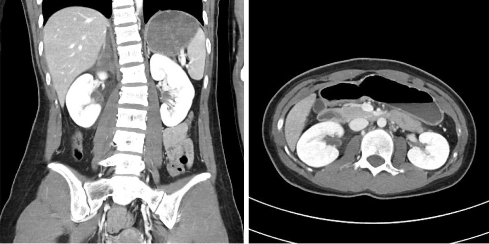
Figure 1 The right adrenal gland was initially thought to be ruptured, with hemorrhage observed in the right perirenal space on a contrast-enhanced abdominopelvic computed tomography scan.
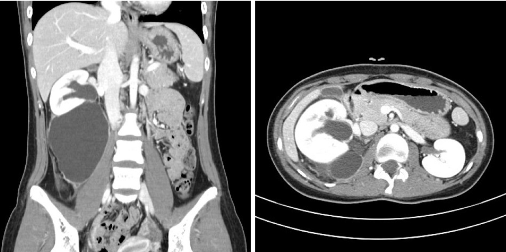
Figure 2 Urine leakage and urinoma with hydronephrosis were observed 3 wk later on a contrast-enhanced abdominopelvic computed tomography scan.
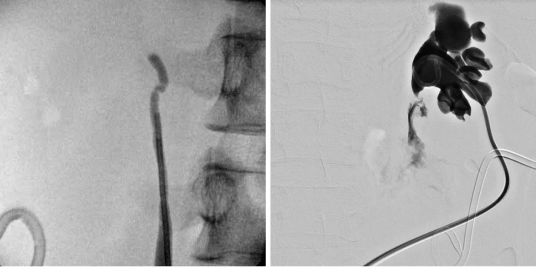
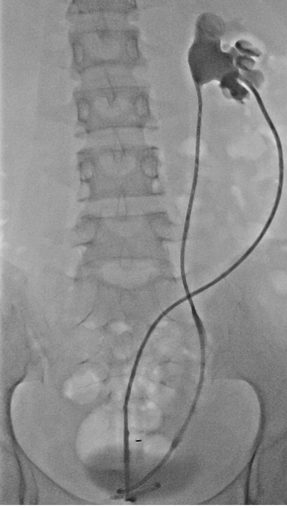
Figure 3 A ureteropelvic junction rupture was revealed by antegrade pyelography.
Discontinuity of the ureter was confirmed by retrograde pyelography and antegrade pyelography.
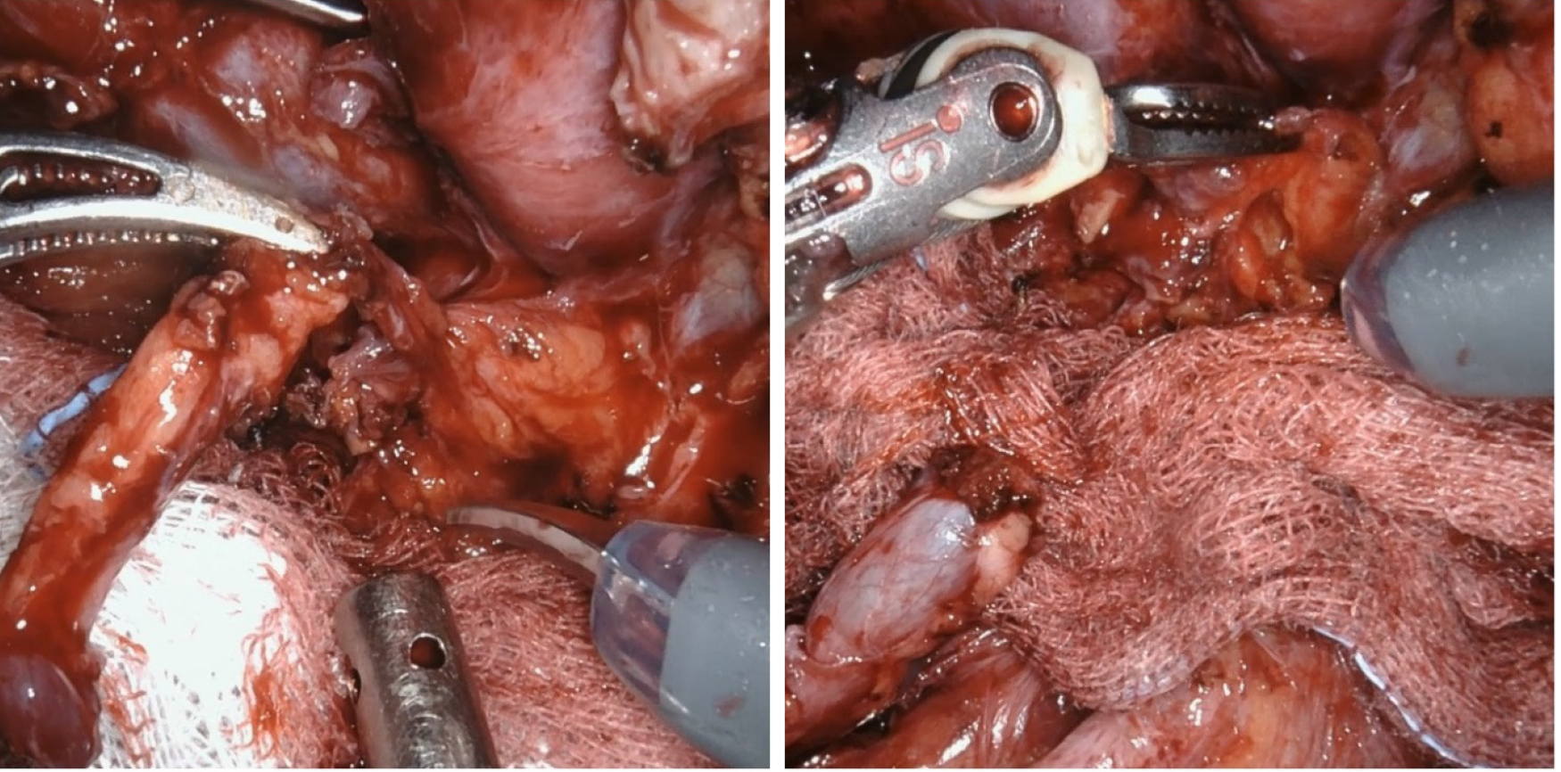
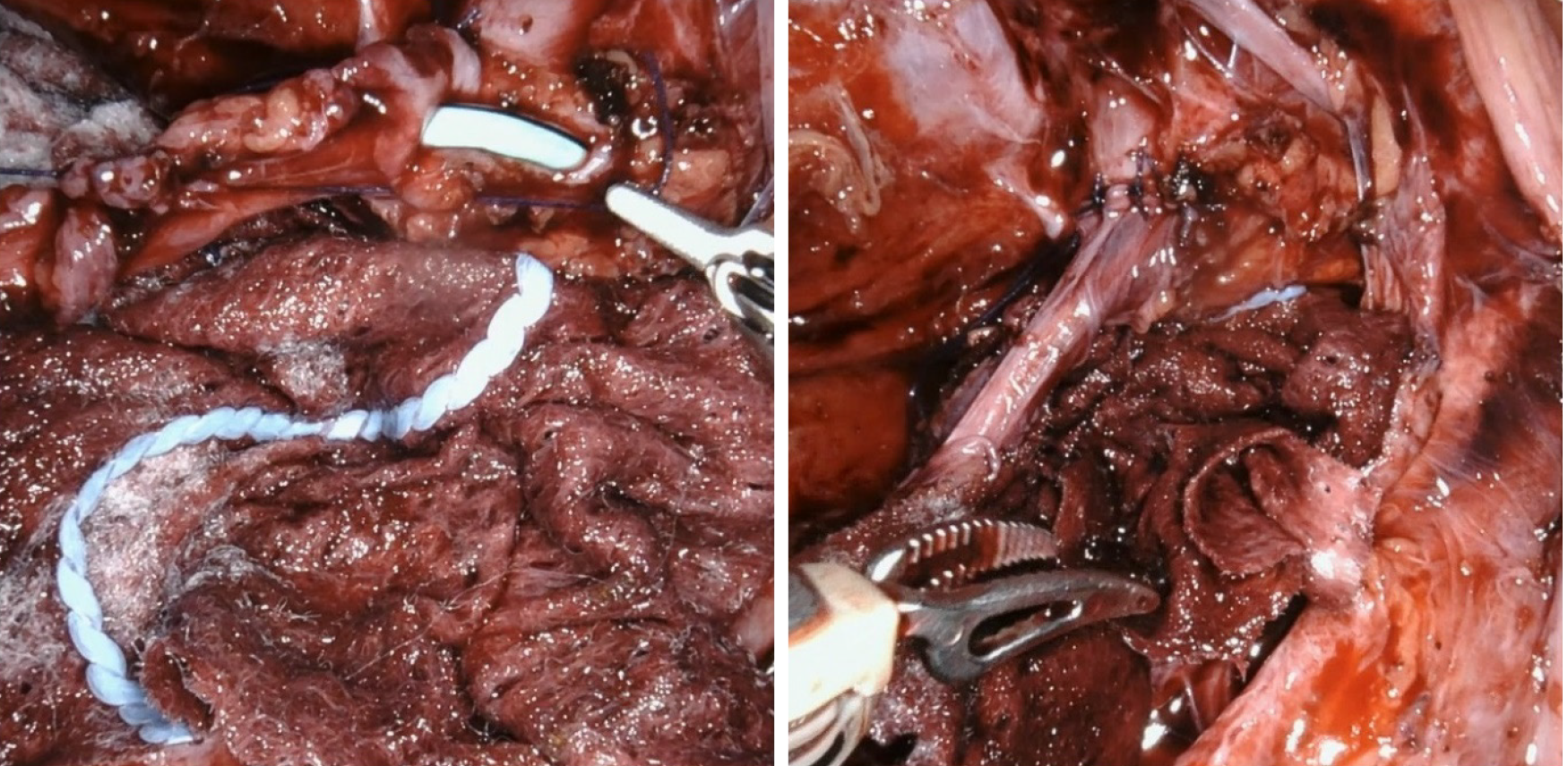
Figure 4 The ureter and renal pelvis were completely separated.
There was no hole in the renal pelvis.
Figure 5 A new incision was made in the renal pelvis to allow anastomosis of the ureter.
Figure 6 Image showing good contrast flow through the stent without leakage.
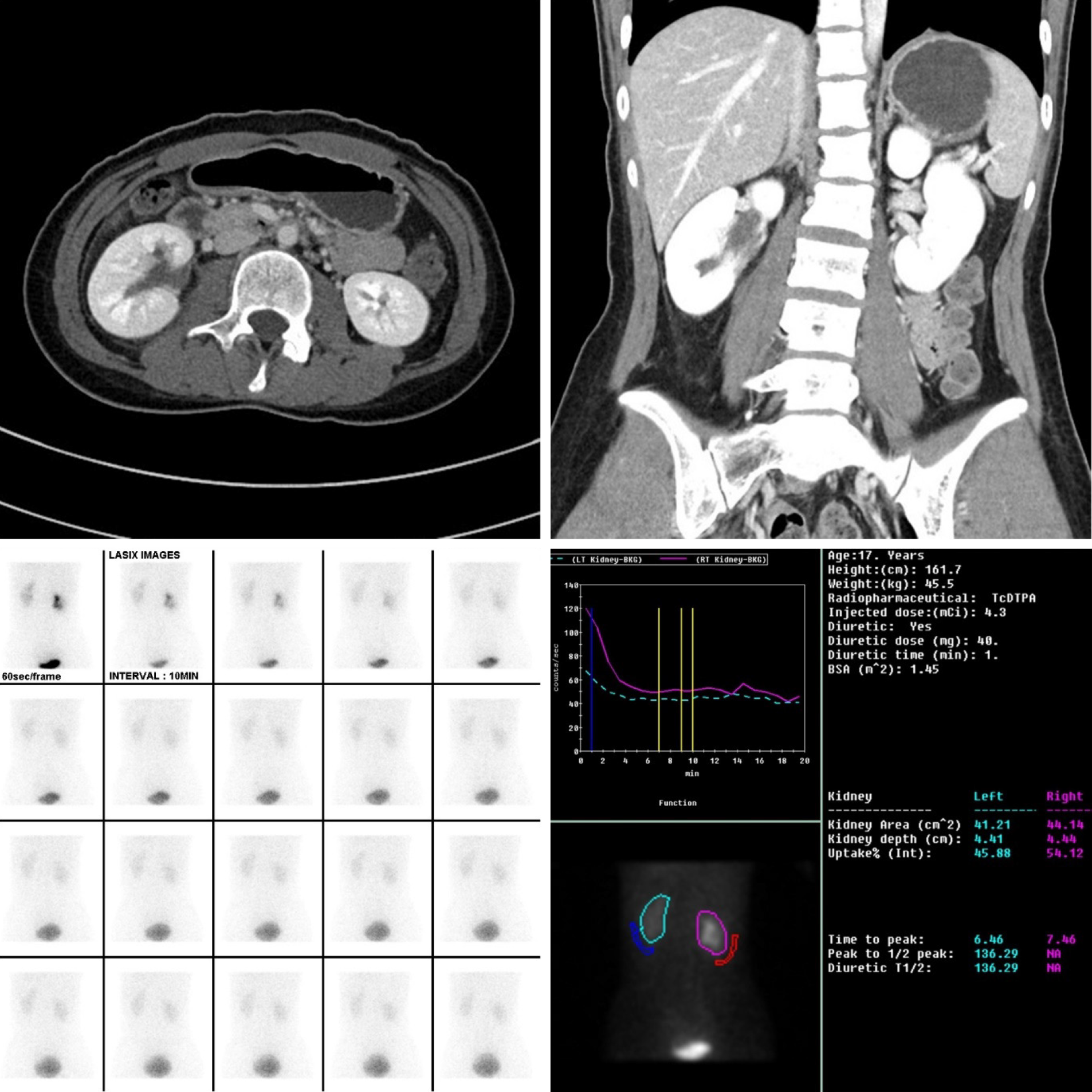
Figure 7 There were no specific findings other than mild renal pelvis dilatation on a contrast-enhanced abdominopelvic computed tomography scan.
Delayed excretion and pelvocalyceal retention of contrast medium in the right kidney, and a rapid response to Furosemide were revealed by renal scintigraphy.
- Citation: Kim SH, Kim WB, Kim JH, Lee SW. Robot-assisted laparoscopic pyeloureterostomy for ureteropelvic junction rupture sustained in a traffic accident: A case report. World J Clin Cases 2020; 8(22): 5802-5808
- URL: https://www.wjgnet.com/2307-8960/full/v8/i22/5802.htm
- DOI: https://dx.doi.org/10.12998/wjcc.v8.i22.5802


