Copyright
©The Author(s) 2020.
World J Clin Cases. Nov 26, 2020; 8(22): 5737-5743
Published online Nov 26, 2020. doi: 10.12998/wjcc.v8.i22.5737
Published online Nov 26, 2020. doi: 10.12998/wjcc.v8.i22.5737
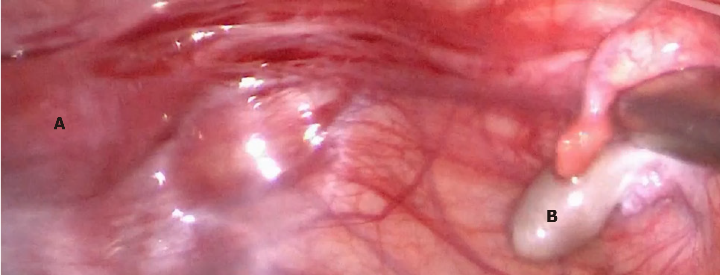
Figure 1 The primordial uterus and right gonad in Case 1.
A: The uterus was approximately 4 cm × 2 cm × 3 cm in size. B: The right gonad looked similar to testicular tissue, and was approximately 1 cm × 1 cm × 0.5 cm in size.
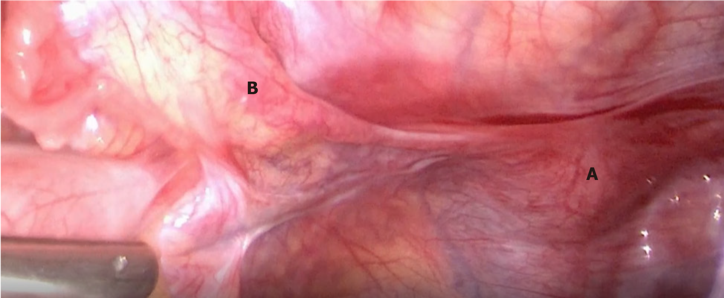
Figure 2 The primordial uterus and the left gonad in Case 1.
A: The uterus; B: The left gonad looked streaky in appearance, and was approximately 0.5 cm × 0.3 cm in size.
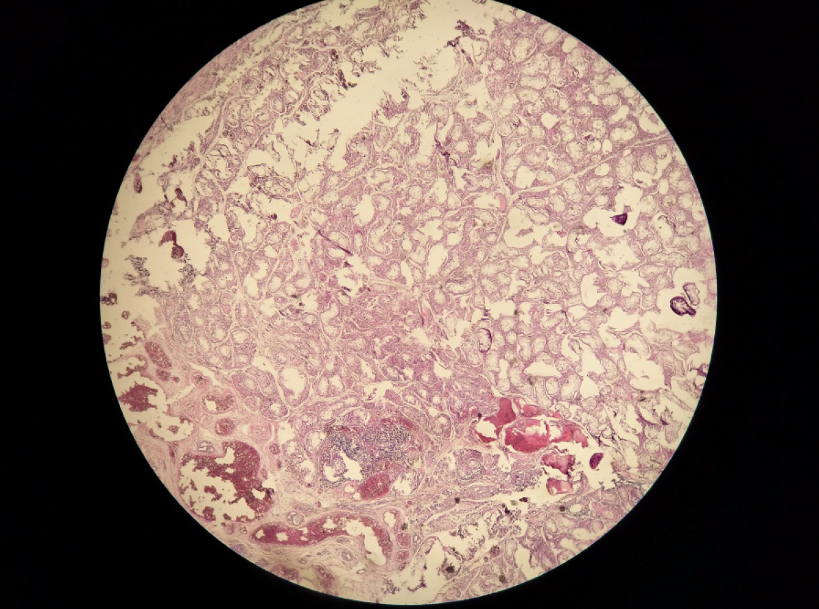
Figure 3 Histopathology of the right gonad.
A: Both tubal epithelium and vas deferens tissue can be seen; B: Sertoli cells could be seen in the seminiferous tubules of the right gonad.
Figure 4 Histopathology of the left gonad in Case 1.
Showing the tubal epithelium, a small amount of hyperplastic fibrous tissue, and smooth muscle tissue, with the presence of angiogenesis.
- Citation: Leng XF, Lei K, Li Y, Tian F, Yao Q, Zheng QM, Chen ZH. Gonadal dysgenesis in Turner syndrome with Y-chromosome mosaicism: Two case reports. World J Clin Cases 2020; 8(22): 5737-5743
- URL: https://www.wjgnet.com/2307-8960/full/v8/i22/5737.htm
- DOI: https://dx.doi.org/10.12998/wjcc.v8.i22.5737


