Copyright
©The Author(s) 2020.
World J Clin Cases. Nov 26, 2020; 8(22): 5715-5721
Published online Nov 26, 2020. doi: 10.12998/wjcc.v8.i22.5715
Published online Nov 26, 2020. doi: 10.12998/wjcc.v8.i22.5715
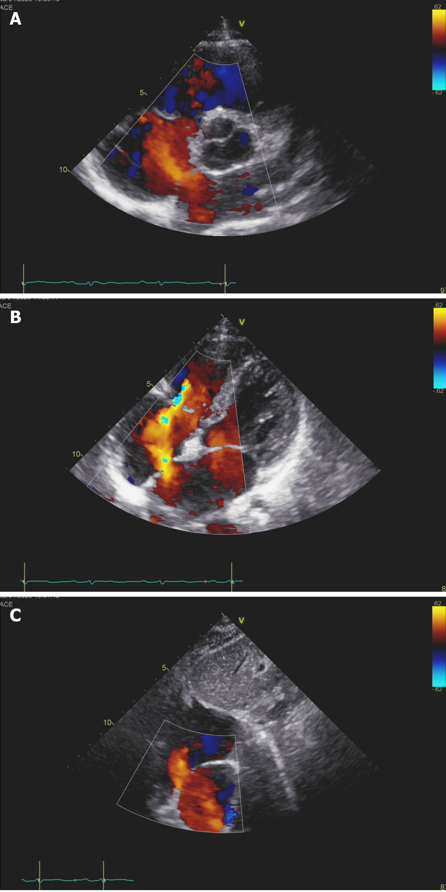
Figure 1 Transthoracic echocardiography showed secundum atrial septal defect with left to right shunt.
A: Aortic root short axis view; B: Parasternal four chamber views; C: Subxiphoid biatrial view.
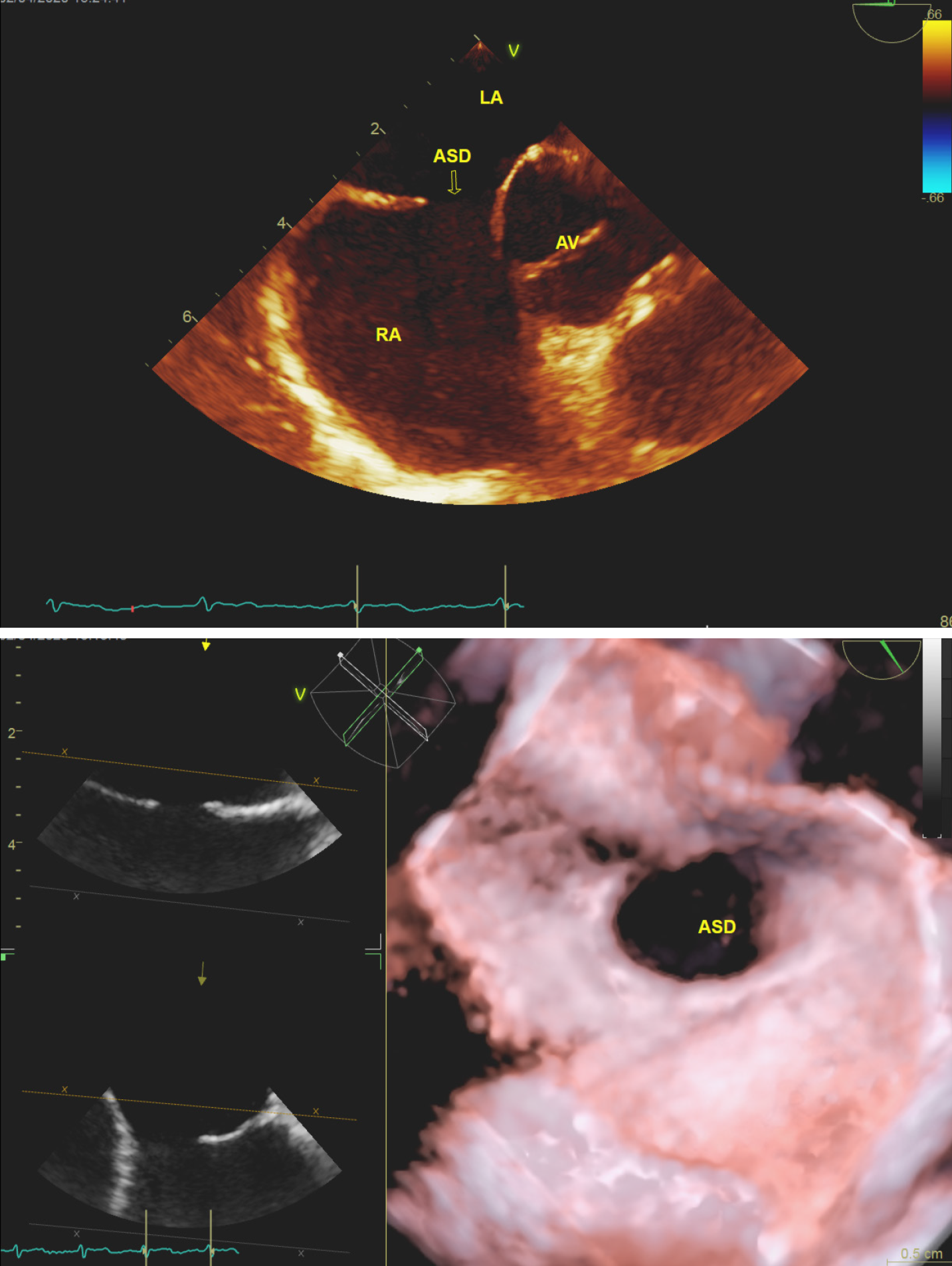
Figure 2 Three-dimensional transthoracic echocardiography showed the atrial septal defect rendered morphology of the atrial septal defect is oval and next to the aortic root.
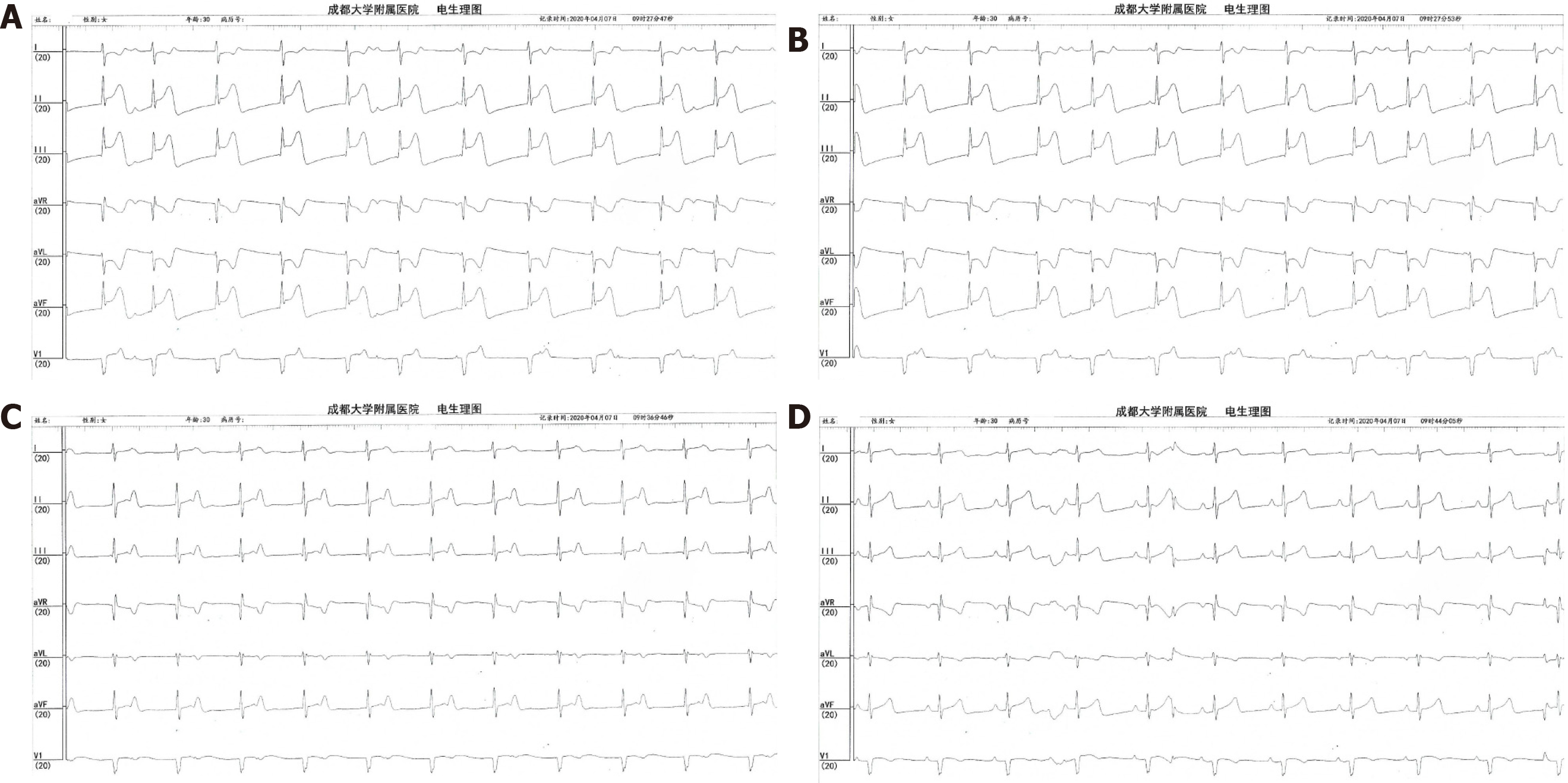
Figure 3 Electrophysiological map.
A: The ST duration elevated when the occluder disc unfolded; B: Completed atrioventricular block; C: The ST duration fell to normal levels, and the atrioventricular block was not recovered when the occluder retreated; D: Sinus rhythm come back about 10 min later.
- Citation: He C, Zhou Y, Tang SS, Luo LH, Feng K. Completed atrioventricular block induced by atrial septal defect occluder unfolding: A case report. World J Clin Cases 2020; 8(22): 5715-5721
- URL: https://www.wjgnet.com/2307-8960/full/v8/i22/5715.htm
- DOI: https://dx.doi.org/10.12998/wjcc.v8.i22.5715


