Copyright
©The Author(s) 2020.
World J Clin Cases. Nov 6, 2020; 8(21): 5409-5414
Published online Nov 6, 2020. doi: 10.12998/wjcc.v8.i21.5409
Published online Nov 6, 2020. doi: 10.12998/wjcc.v8.i21.5409
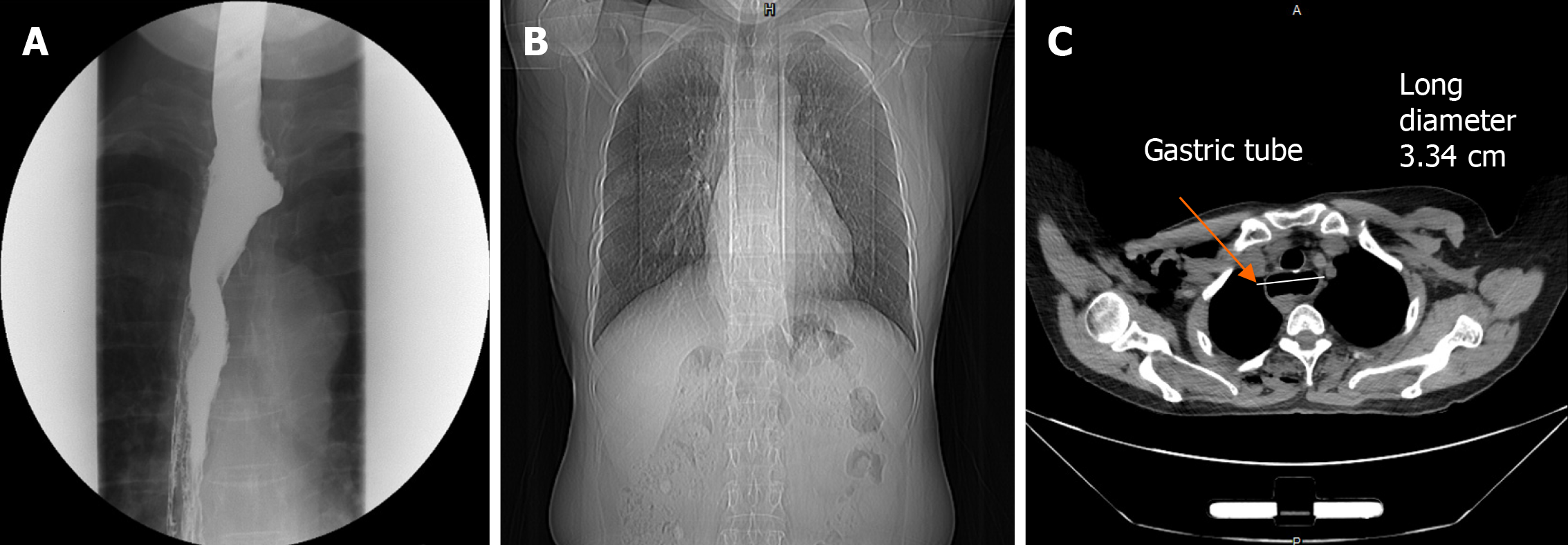
Figure 1 Preoperative examination.
A: Preoperative esophagography showed patency of the reconstructed esophagus and no local fistula; B: Preoperative chest computed tomography showed right lung lower lobe nodules with no active lung lesion; C: Preoperative chest computed tomography showed gastric tube mild dilatation.
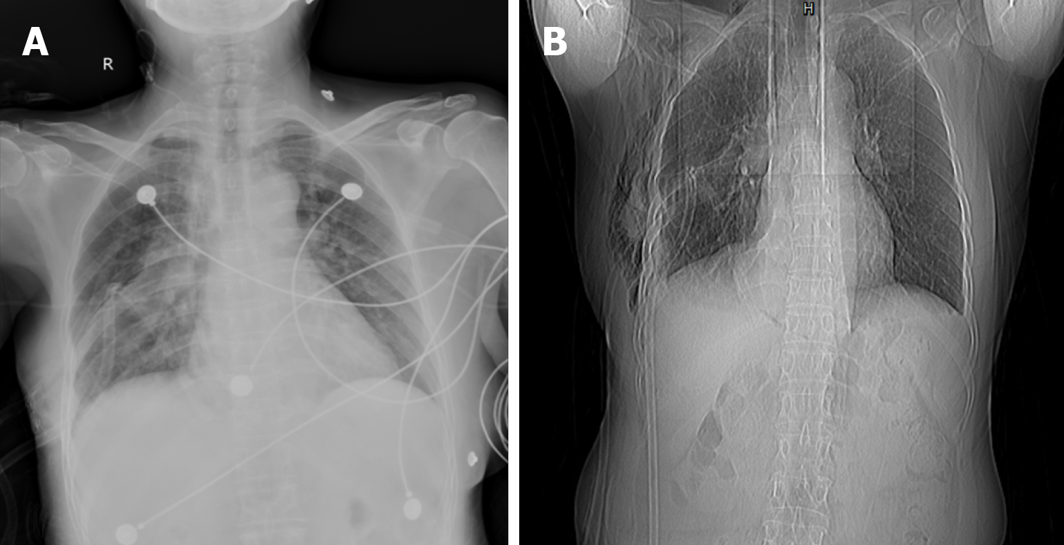
Figure 2 Postoperative examination.
A: Immediate postoperative chest X-ray showed ill-defined frosted hyaline shadow with exudative lesions in both lungs; B: At postoperative day 6, follow-up chest X-ray showed exudative lesions in both lungs were markedly reduced.
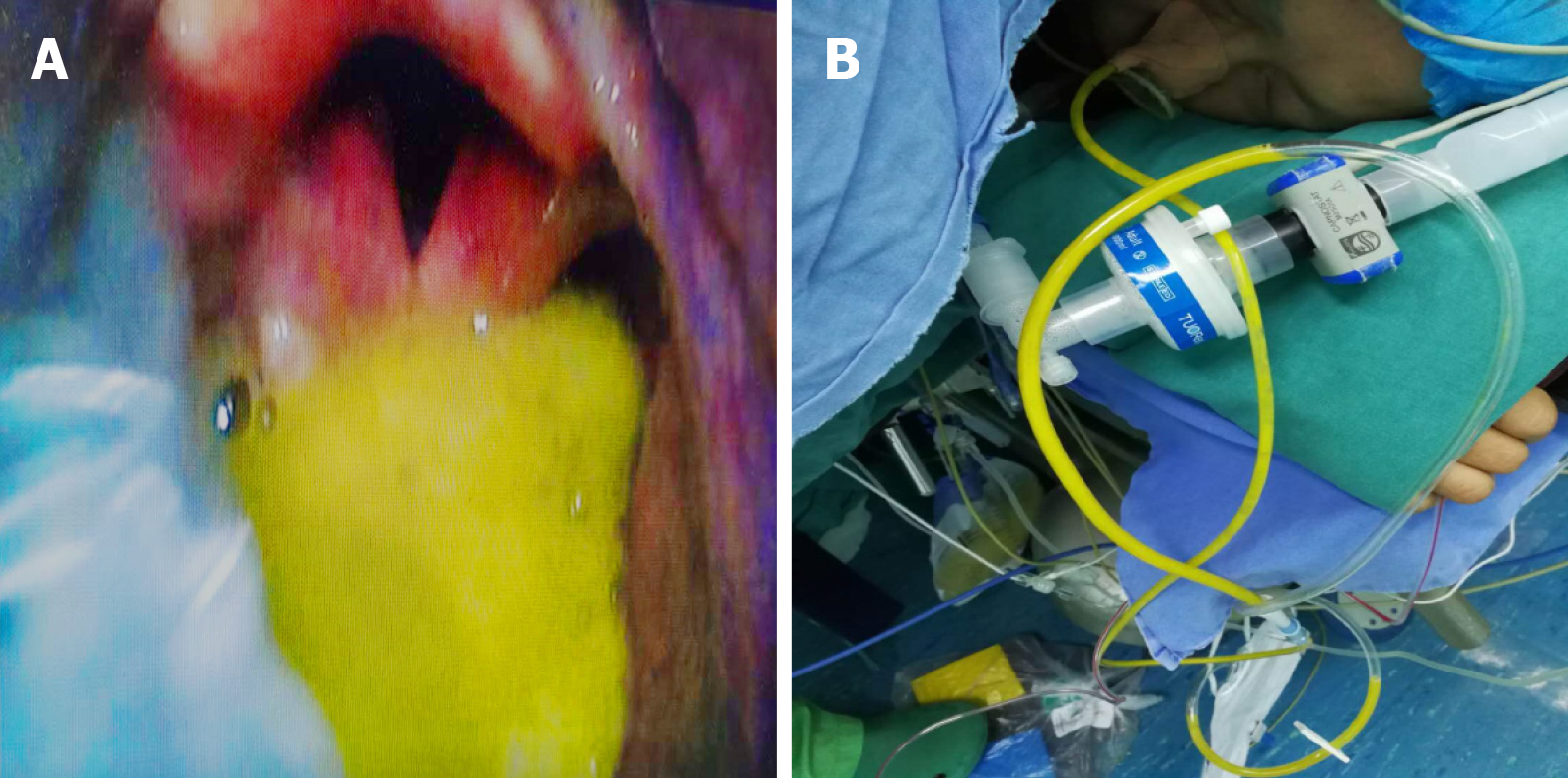
Figure 3 A video laryngoscope.
A: Reflux of yellow–green gastric fluid into the pharyngeal cavity; B: Nasogastric tube to drain approximately 100 mL of yellow–green liquid.
- Citation: Tang JX, Wang L, Nian WQ, Tang WY, Xiao JY, Tang XX, Liu HL. Aspiration pneumonia during general anesthesia induction after esophagectomy: A case report. World J Clin Cases 2020; 8(21): 5409-5414
- URL: https://www.wjgnet.com/2307-8960/full/v8/i21/5409.htm
- DOI: https://dx.doi.org/10.12998/wjcc.v8.i21.5409


