Copyright
©The Author(s) 2020.
World J Clin Cases. Nov 6, 2020; 8(21): 5401-5408
Published online Nov 6, 2020. doi: 10.12998/wjcc.v8.i21.5401
Published online Nov 6, 2020. doi: 10.12998/wjcc.v8.i21.5401
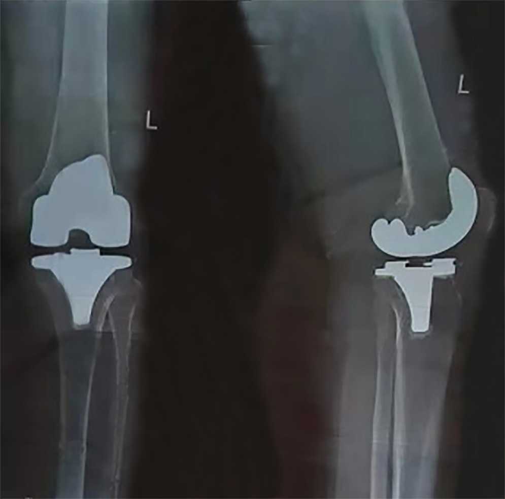
Figure 1 Anteroposterior and lateral radiographs after primary total knee arthroplasty.
The surgery was performed 20 mo prior to current presentation.
Figure 2 Radiograph of the patient’s left knee after prosthesis removal and insertion of an antibiotic (vancomycin) impregnated cement spacer.
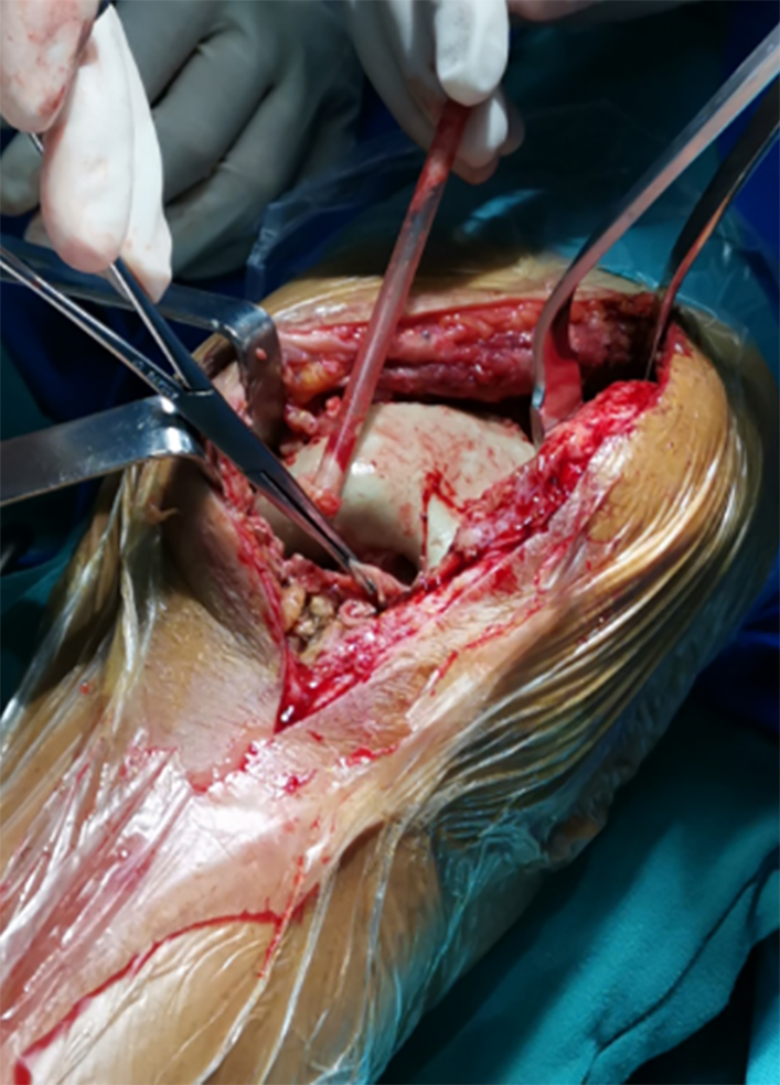
Figure 3 Cheesy like necrotic tissue was observed on the surface of the spacer which had an unpleasant odor during the second debridement procedure.
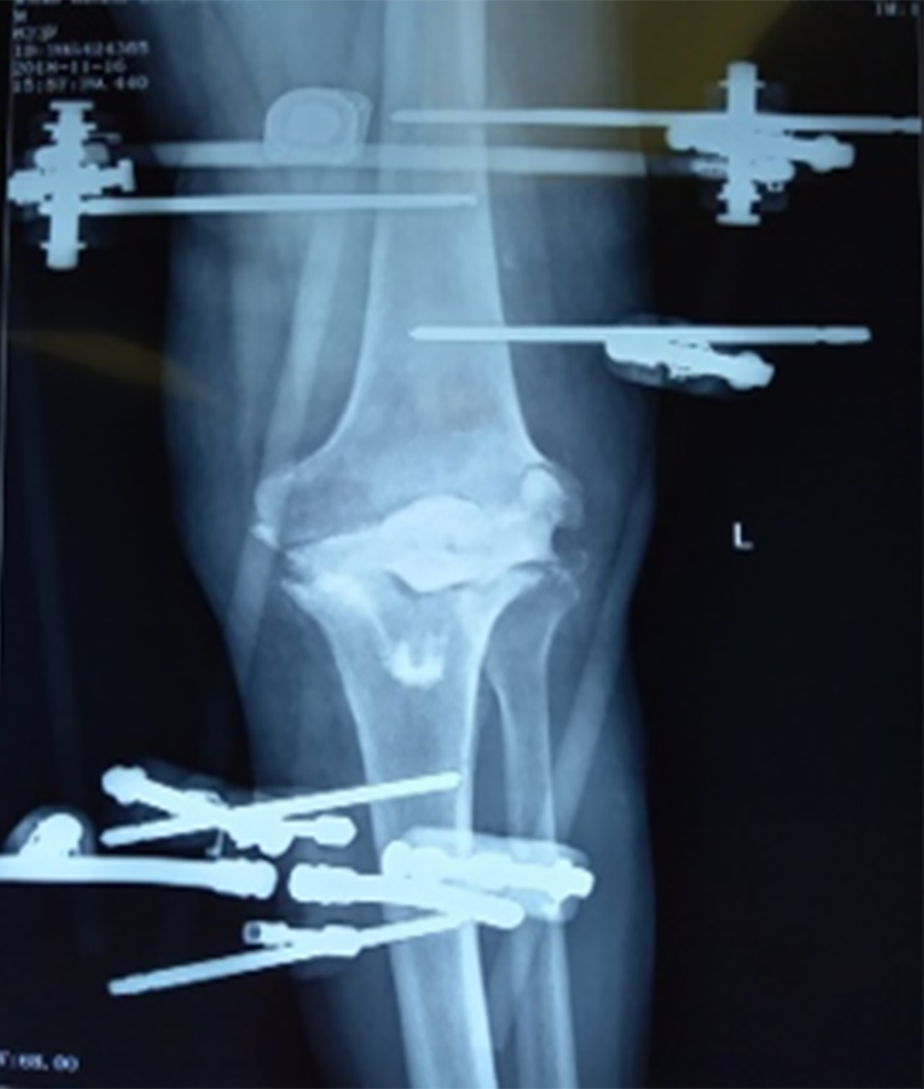
Figure 4 Radiograph of the patient’s left knee after the second debridement.
Figure 5 Radiograph of the left intertrochanteric fracture (31A2.
2) after the second debridement.
Figure 6 Ipsilateral intertrochanteric fracture fixed by proximal femoral nail anterotation.
Figure 7 Knee arthrodesis with autograft using a double-plate fixation.
Figure 8 Radiography at the two-year follow-up.
- Citation: Xin J, Guo QS, Zhang HY, Zhang ZY, Talmy T, Han YZ, Xie Y, Zhong Q, Zhou SR, Li Y. Candidal periprosthetic joint infection after primary total knee arthroplasty combined with ipsilateral intertrochanteric fracture: A case report. World J Clin Cases 2020; 8(21): 5401-5408
- URL: https://www.wjgnet.com/2307-8960/full/v8/i21/5401.htm
- DOI: https://dx.doi.org/10.12998/wjcc.v8.i21.5401


