Copyright
©The Author(s) 2020.
World J Clin Cases. Nov 6, 2020; 8(21): 5380-5388
Published online Nov 6, 2020. doi: 10.12998/wjcc.v8.i21.5380
Published online Nov 6, 2020. doi: 10.12998/wjcc.v8.i21.5380
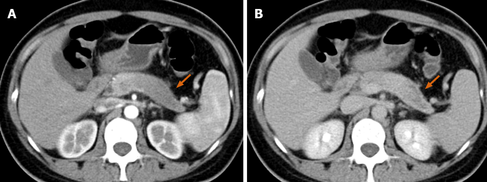
Figure 1 Change in the contour of the pancreatic body and tail, with the effacement of lobules.
A: The lesion is hypo-enhanced in the pancreatic parenchyma phase (orange arrow), with progressive delayed enhancement; B: Internal necrosis is seen in the delayed phase.
Figure 2 Bilateral cystic masses in the pelvis alongside the uterus.
The masses measure 15.1 cm × 12.0 cm and 5.7 cm × 4.3 cm (A and B, respectively). Both are thin-walled with multiple enhancing septa (orange arrow). There is no evidence of liver or peritoneal metastases.
Figure 3 Specimens of the ovarian mass and distal pancreas with spleen.
A: An ovarian metastatic mass with a cystic-solid profile contains many mucinous cysts; B: A pale solid mass sized 4 cm × 3.5 cm is seen at the tail of the pancreas.
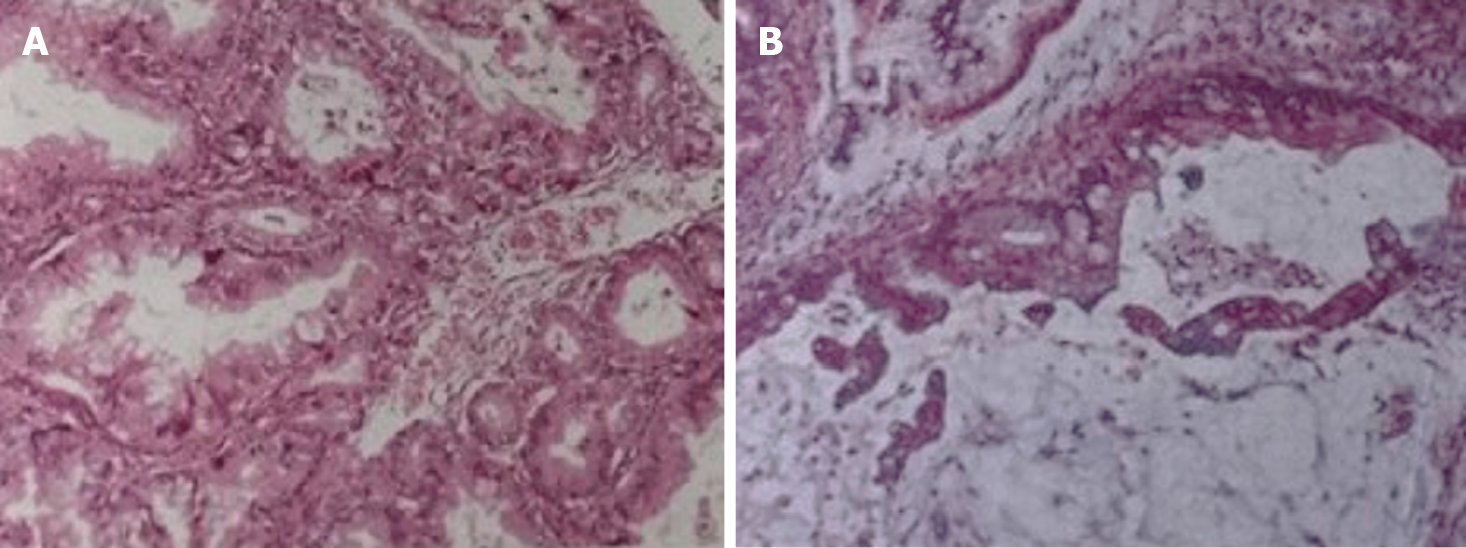
Figure 4 Histopathological analysis of the resected tumors.
A: Pancreatic tumor; B: Ovarian mass.
- Citation: Wang SD, Zhu L, Wu HW, Dai MH, Zhao YP. Pancreatic cancer with ovarian metastases: A case report and review of the literature. World J Clin Cases 2020; 8(21): 5380-5388
- URL: https://www.wjgnet.com/2307-8960/full/v8/i21/5380.htm
- DOI: https://dx.doi.org/10.12998/wjcc.v8.i21.5380


