Copyright
©The Author(s) 2020.
World J Clin Cases. Nov 6, 2020; 8(21): 5304-5312
Published online Nov 6, 2020. doi: 10.12998/wjcc.v8.i21.5304
Published online Nov 6, 2020. doi: 10.12998/wjcc.v8.i21.5304
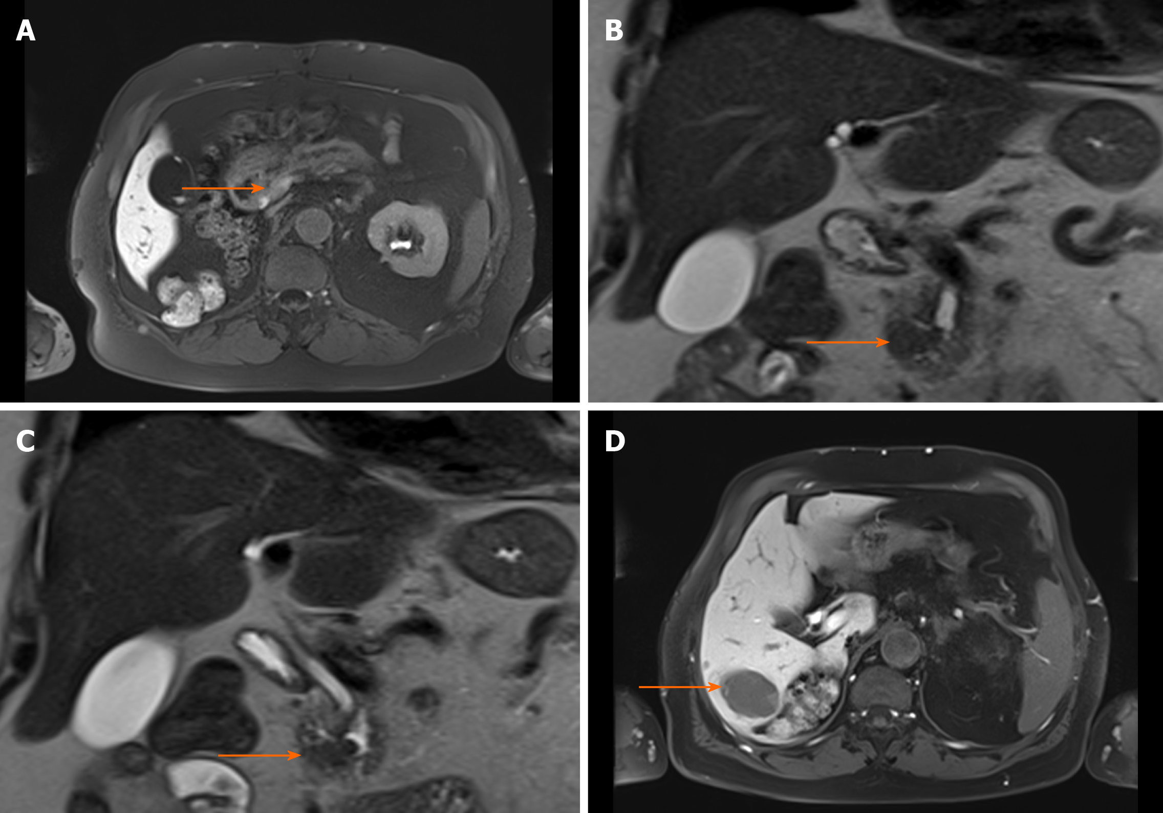
Figure 1 T2-weighted magnetic resonance images showing acinar cell carcinoma of the pancreatic head with synchronous liver metastases before surgery.
The patient’s presurgical bloodwork showed lipase of 5580 U/L, amylase of 172 U/L, C-reactive protein of 3.3 mg/dL, and leukocytes of 5 G/L. A: Primary tumor localized in the processus uncinatus; B and C: T2-weighted HASTE cor showing prepapillary tumor (arrow) infiltrating the common hepatic duct, which is extended; D: Hepatic metastasis in liver segment VI (arrow).
Figure 2 Computed tomography at 9 mo after surgery showing hepatic progression of the disease.
The patient’s bloodwork showed lipase of 2384 U/L, amylase of 61 U/L, C-reactive protein of 13.1 mg/dL, and leukocytes of 4.98 G/L. A and B: Multifocal filiae of the liver with hypovascular metastases (arrows), and tumor infiltration of the abdominal wall dorsally of the liver. Of note, compared with pancreatic ductal adenocarcinoma[48], the tumor density of the acinar cell carcinoma in the non-contrast phase and time attenuation curve pattern are different but both tumor entities tend to be hypodense in the contrast-enhanced phases; C: Portal venous phase of recurrent metastasis with solid and cystic properties (liver segment VII).
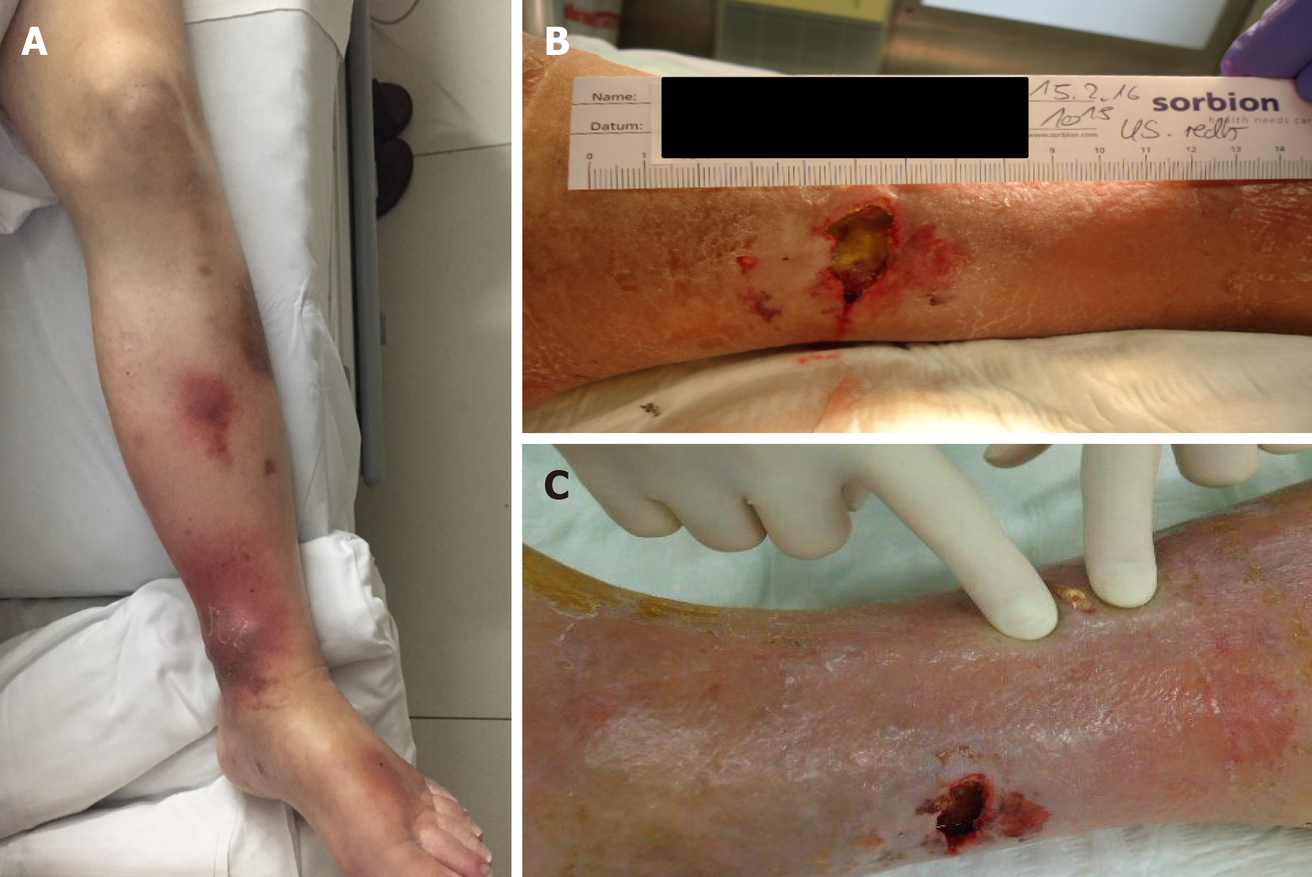
Figure 3 Progressive panniculitis with fat necrosis in the lower extremities at 10 mo after surgery.
The patient’s bloodwork showed lipase of 8414 U/L, amylase of 82 U/L, C-reactive protein of 5.2 mg/dL, and leukocytes of 5.41 G/L. A-C: Erythema suspicious for panniculitis occurred in both legs.
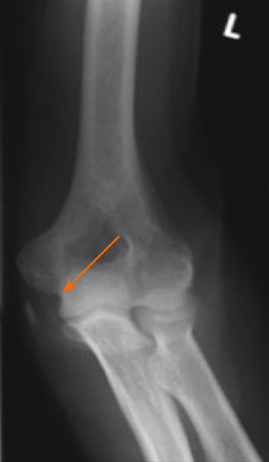
Figure 4 X-ray at 12 mo after surgery showing acute arthritis of the left elbow with epicondylitis (arrow).
The patient’s bloodwork showed lipase of 13410 U/L, amylase of 22 U/L, C-reactive protein of 22.3 mg/dL, and leukocytes of 10.2 G/L.
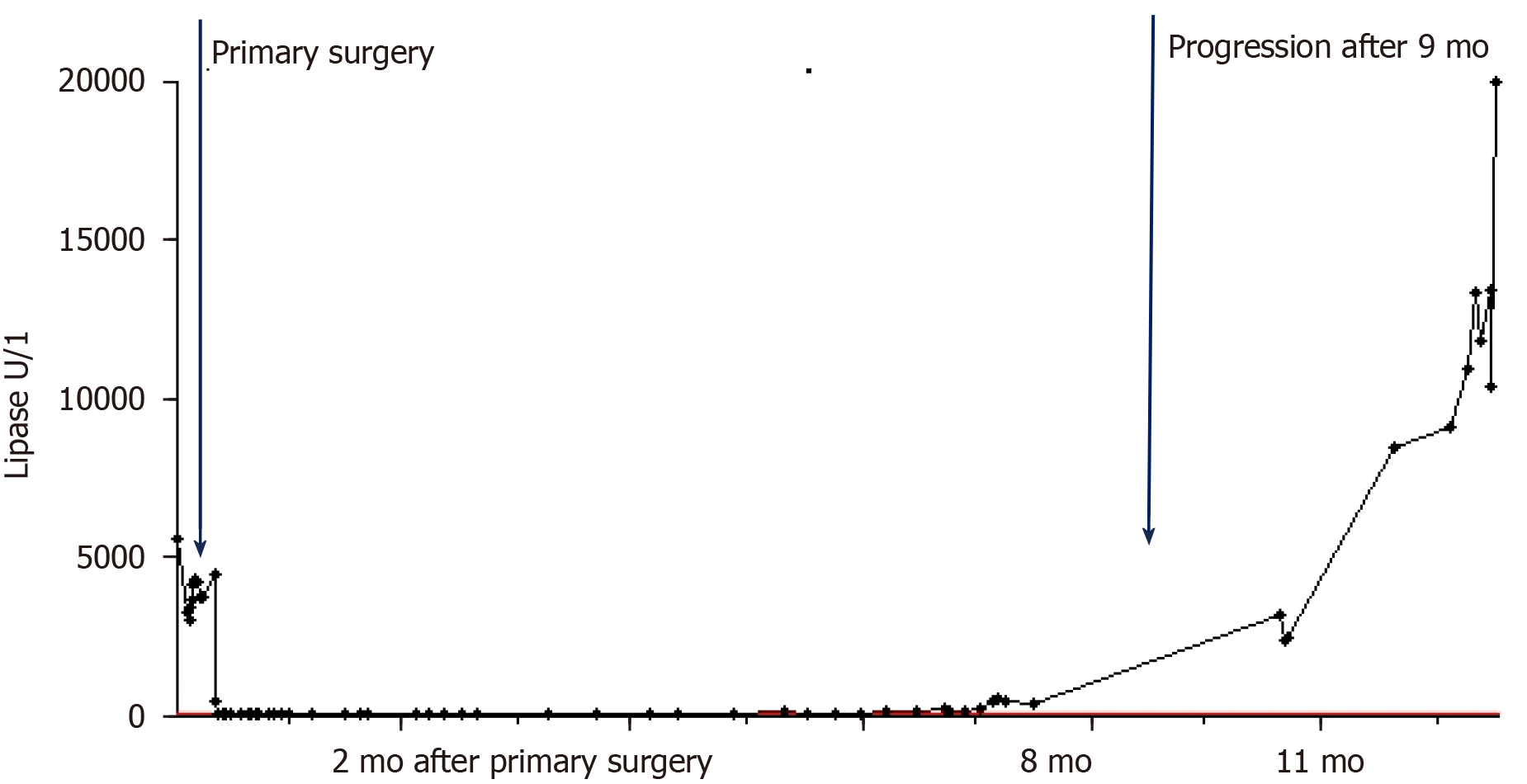
Figure 5 Dynamics of serum lipase levels starting before surgery and during follow-up.
The maximal concentration of lipase was 19940 U/L, of C-reactive protein was 22.9 mg/dL, and leukocytes was 11.2 G/L. The carcinoembryonic antigen (CEA) an CA19-9 tumor marker levels were collected during follow-up as well: CEA 1.5 ng/mL and CA19-9 13.3 U/mL (first surgery), CEA 1.7 ng/mL and CA19-9 10.2 U/mL (6 mo after primary surgery), CEA 1.7 ng/mL and CA19-9 9.9 U/mL (9 mo after primary surgery).
- Citation: Miksch RC, Schiergens TS, Weniger M, Ilmer M, Kazmierczak PM, Guba MO, Angele MK, Werner J, D'Haese JG. Pancreatic panniculitis and elevated serum lipase in metastasized acinar cell carcinoma of the pancreas: A case report and review of literature. World J Clin Cases 2020; 8(21): 5304-5312
- URL: https://www.wjgnet.com/2307-8960/full/v8/i21/5304.htm
- DOI: https://dx.doi.org/10.12998/wjcc.v8.i21.5304


