Copyright
©The Author(s) 2020.
World J Clin Cases. Oct 26, 2020; 8(20): 5049-5056
Published online Oct 26, 2020. doi: 10.12998/wjcc.v8.i20.5049
Published online Oct 26, 2020. doi: 10.12998/wjcc.v8.i20.5049
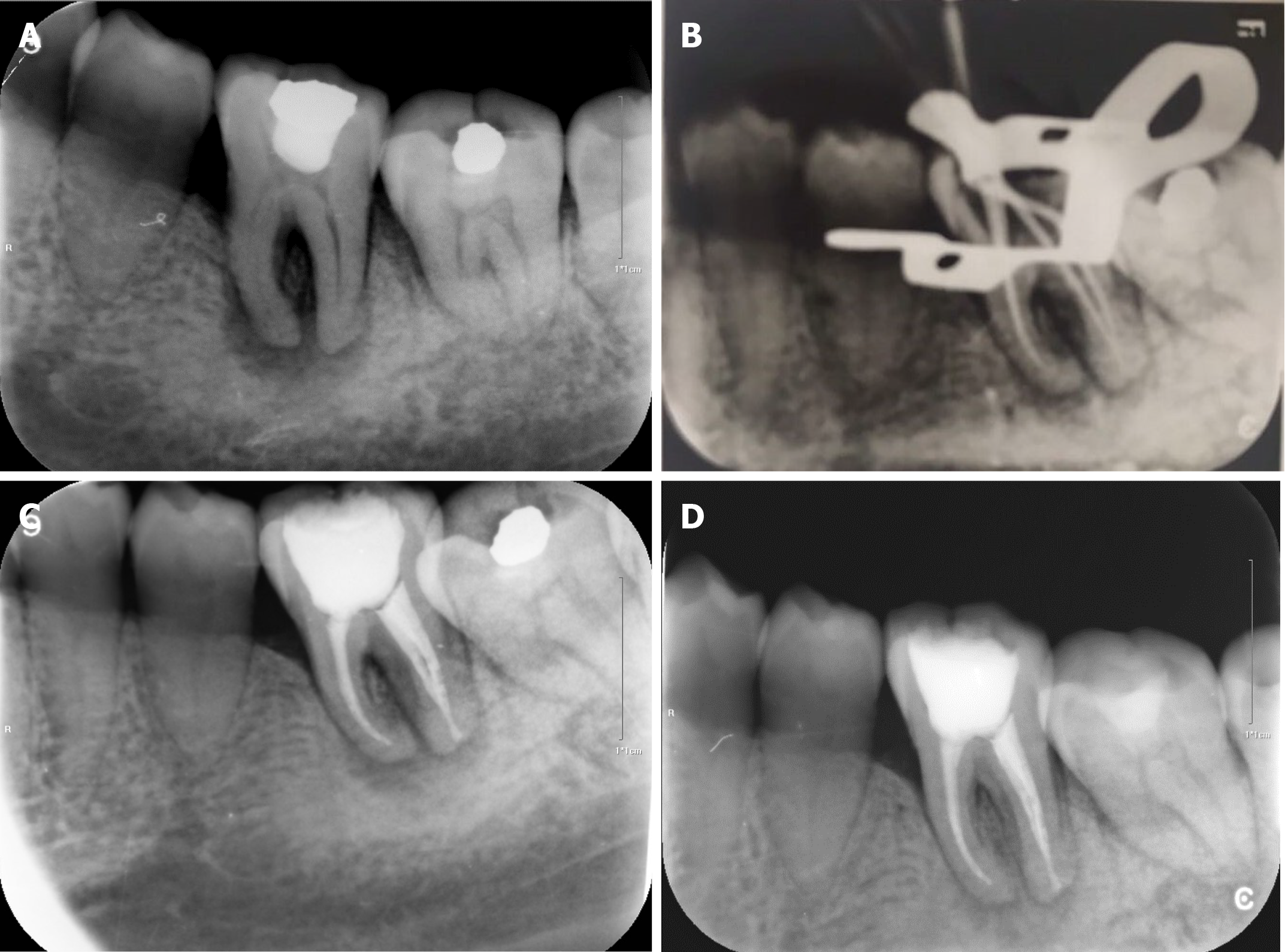
Figure 1 Periapical radiograph.
A: Initial preoperative periapical radiograph; B: Gutta-Percha cone fitting and working length confirmation with periapical radiograph; C: Post-obturation periapical radiograph; D: Periapical radiograph at the 3 mo follow-up.
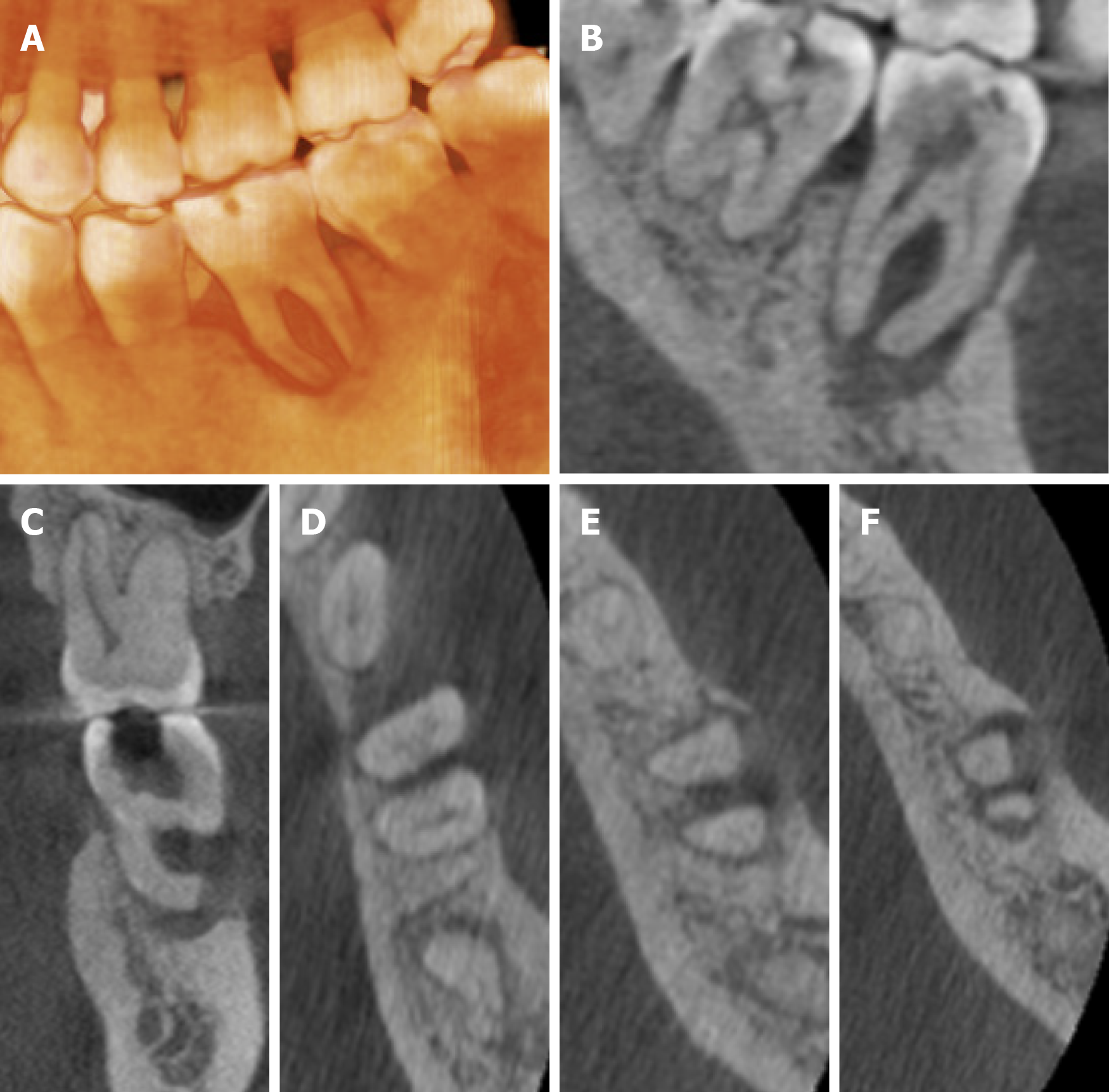
Figure 2 Images of cone beam computed tomography showing buccal, bifurcation, and apical bone resorption (A-F).
Figure 3 Flow chart timeline of the treatment plan.
- Citation: Alshawwa H, Wang JF, Liu M, Sun SF. Successful management of a tooth with endodontic-periodontal lesion: A case report. World J Clin Cases 2020; 8(20): 5049-5056
- URL: https://www.wjgnet.com/2307-8960/full/v8/i20/5049.htm
- DOI: https://dx.doi.org/10.12998/wjcc.v8.i20.5049


