Copyright
©The Author(s) 2020.
World J Clin Cases. Oct 26, 2020; 8(20): 4895-4901
Published online Oct 26, 2020. doi: 10.12998/wjcc.v8.i20.4895
Published online Oct 26, 2020. doi: 10.12998/wjcc.v8.i20.4895
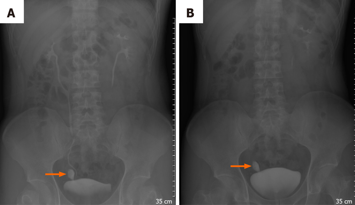
Figure 1 Intravenous pyelography was further investigated, and on 5-min film a well-defined contrast shape structure was observed; at 30-min film, the contrast was still trapped insides.
Differential diagnosis could be bladder rupture, ureteral tear, or anatomical abnormality. A: 5-min film; and B: 30-min film.
Figure 2 The coronal and axial of computed tomography urogram clearly demonstrated the anatomical relation of the Hutch diverticulum with the right ureter and how it goes into the urinary bladder.
A: Coronal; and B: Axial.
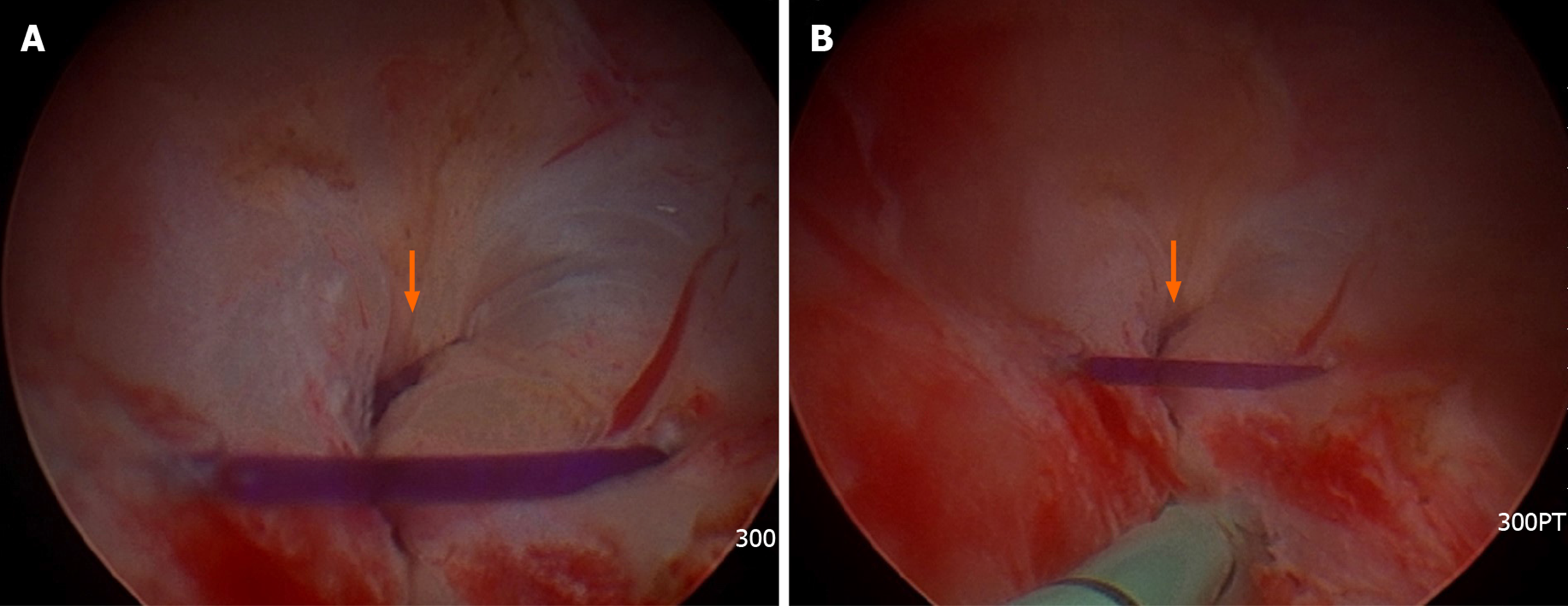
Figure 3 The right ureteral orifice under the cystoscopy.
A: The opening the diverticulum was obviously visible on the superolateral side of the right ureteral orifice under the cystoscopy; and B: We left a ureteral catheter along the right ureter to relieve the hydronephrosis temporarily.
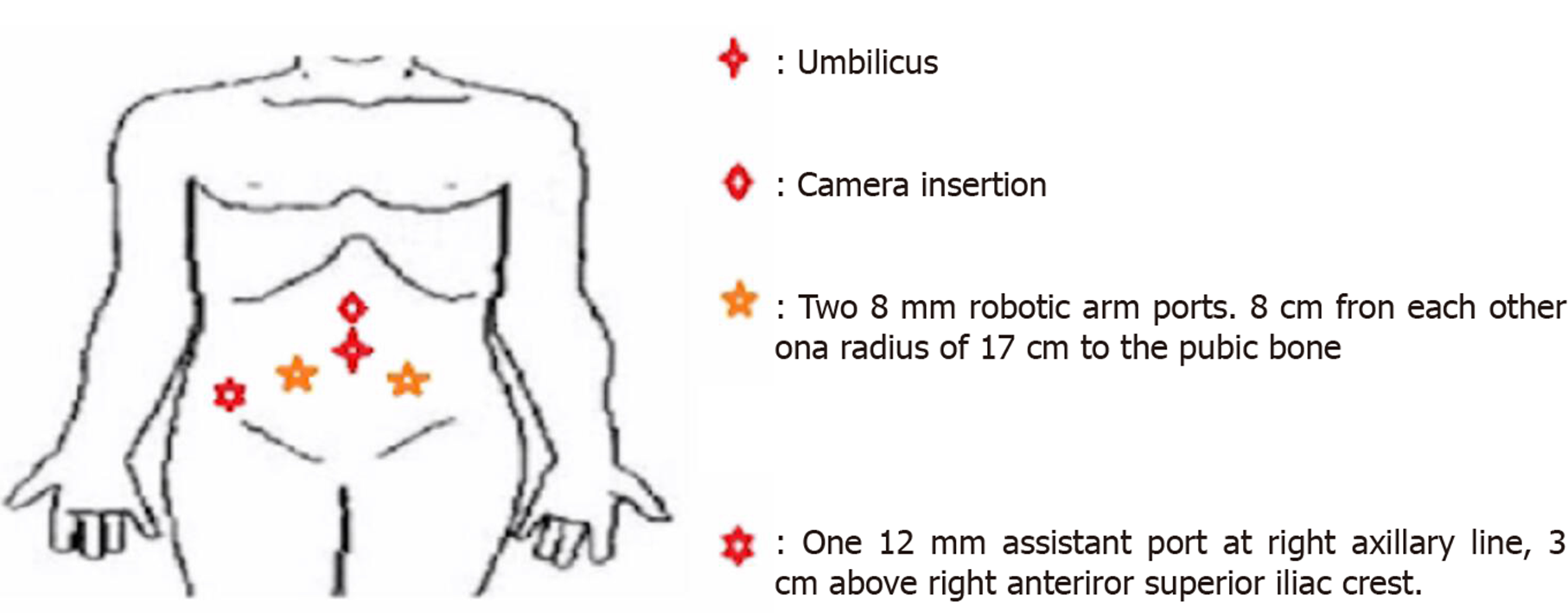
Figure 4 Ports insertion of robotic-assisted diverticulectomy and reconstruction.
Figure 5 The 3-cm width hutch diverticulum was bulged out by saline solution instillation at first before the operation.
A: After going into the cul-de-sac by transperitoneal approach, it could be located by dissecting lateral to the right seminal vesicle; B: We carefully dissected between the ureter and the diverticulum; and C: Cutting it off from the neck, reconstruction was performed by the running suture.
Figure 6 Postoperative follow-up at the second week after surgery was arranged.
Under ultrasound, right hydronephrosis was relieved A: Preoperative hydronephrosis; B: Postoperative dissolution of hydronephrosis; and C: No extravasation was seen at the bladder and ureteral reconstruction.
- Citation: Yang CH, Lin YS, Ou YC, Weng WC, Huang LH, Lu CH, Hsu CY, Tung MC. Adult metaplastic hutch diverticulum with robotic-assisted diverticulectomy and reconstruction: A case report. World J Clin Cases 2020; 8(20): 4895-4901
- URL: https://www.wjgnet.com/2307-8960/full/v8/i20/4895.htm
- DOI: https://dx.doi.org/10.12998/wjcc.v8.i20.4895


