Copyright
©The Author(s) 2020.
World J Clin Cases. Jan 26, 2020; 8(2): 464-470
Published online Jan 26, 2020. doi: 10.12998/wjcc.v8.i2.464
Published online Jan 26, 2020. doi: 10.12998/wjcc.v8.i2.464
Figure 1 Enhanced computed tomography imaging.
A: Thickening of the gallbladder wall and irregular density soft tissue extending from the neck and invading into the cavity and intrahepatic bile ducts with intrahepatic bile duct dilatation by approximately 2.5 cm × 1.3 cm; B: The cystic duct lumen was narrowed and a circular calcification-like high-density shadow was visualized at the end of the common bile duct at approximately 0.8 cm.
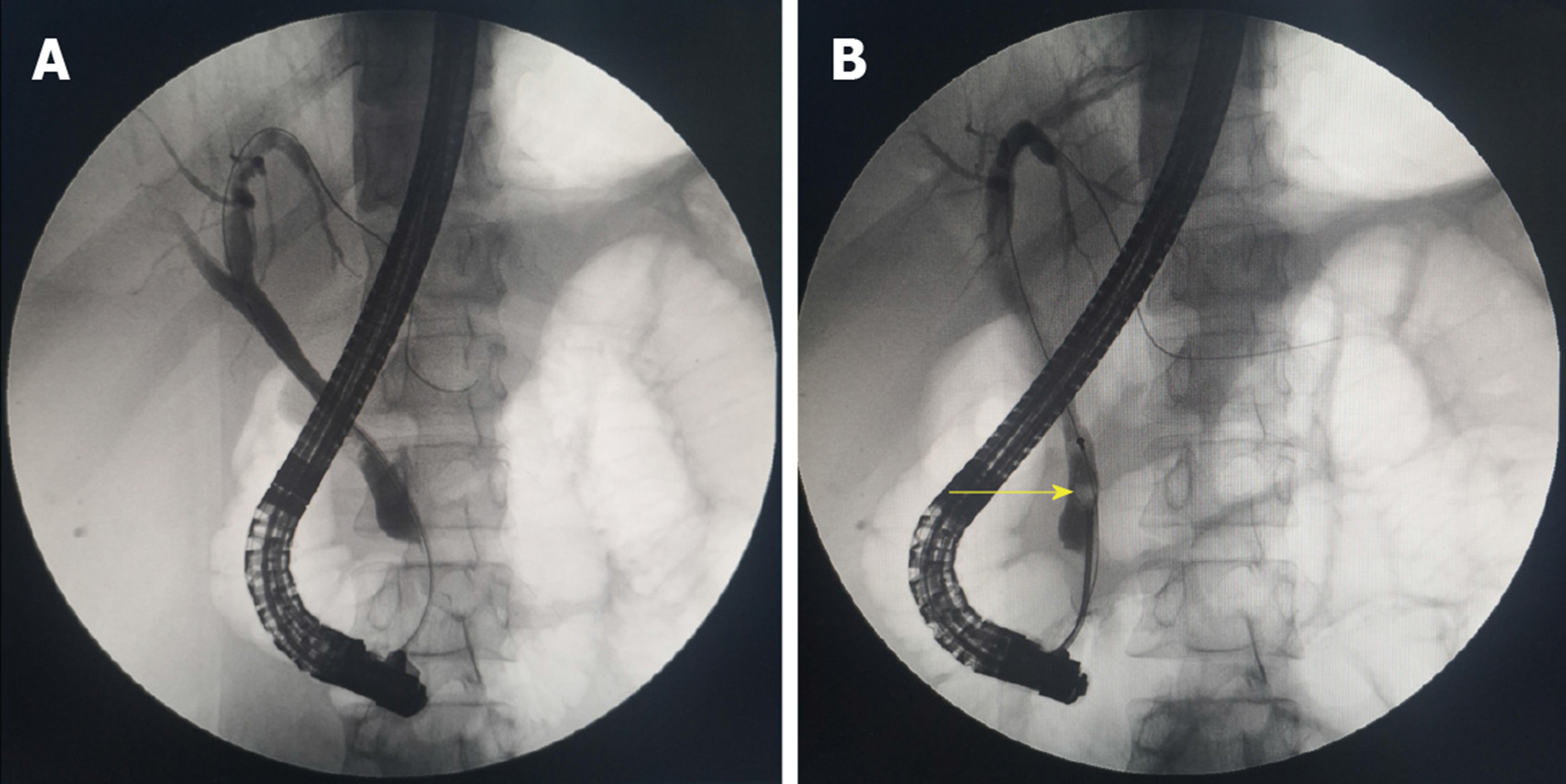
Figure 2 Endoscopic retrograde cholangio-pancreatography.
A: Bile duct dilatation; B: Gallstones at the end of the common bile duct.
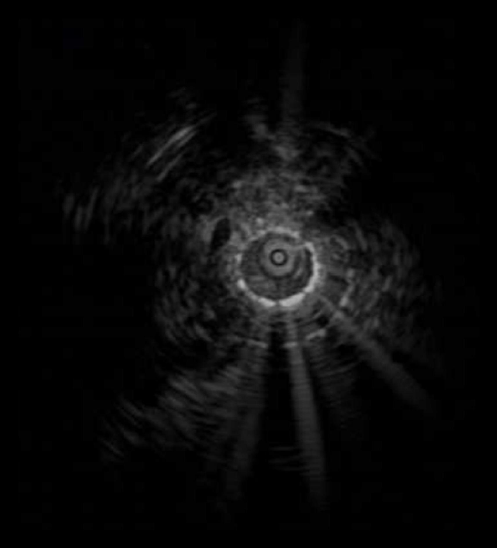
Figure 3 Intraductal ultrasound imaging.
Homogeneous hyperechoic lesions with smooth margins of benign bile duct stricture were visualized.
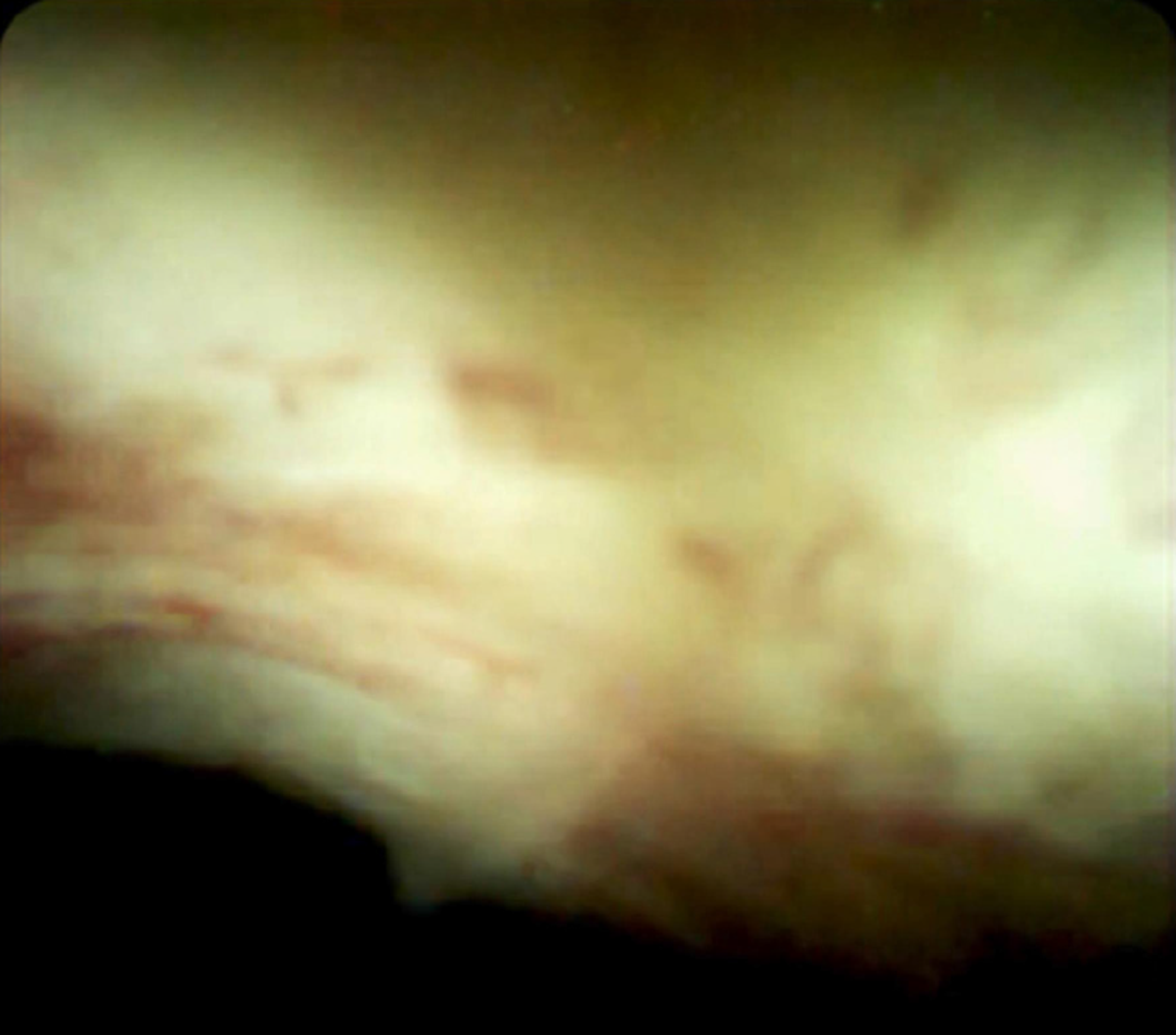
Figure 4 SpyGlass Direct Visualization System imaging.
Inflammatory change in the bile duct mucosa was observed.
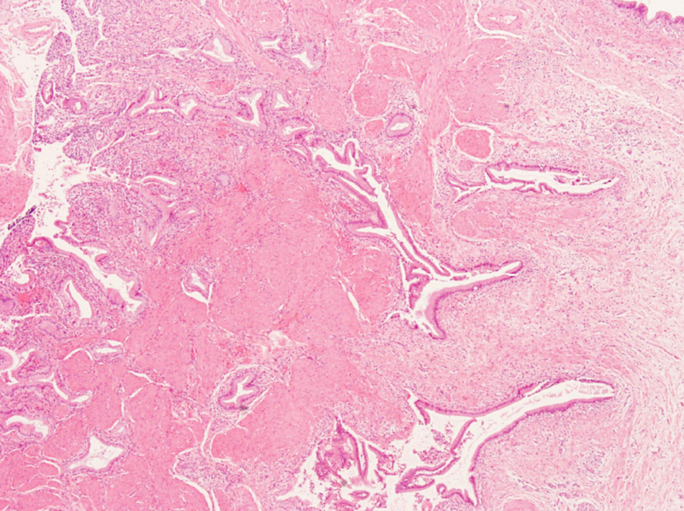
Figure 5 Pathological examination.
Gallbladder adenomyomatosis with chronic cholecystitis and acute suppurative inflammation was present without signs of gallbladder carcinomas at the incisal margin of the liver and neck of the gallbladder (×40).
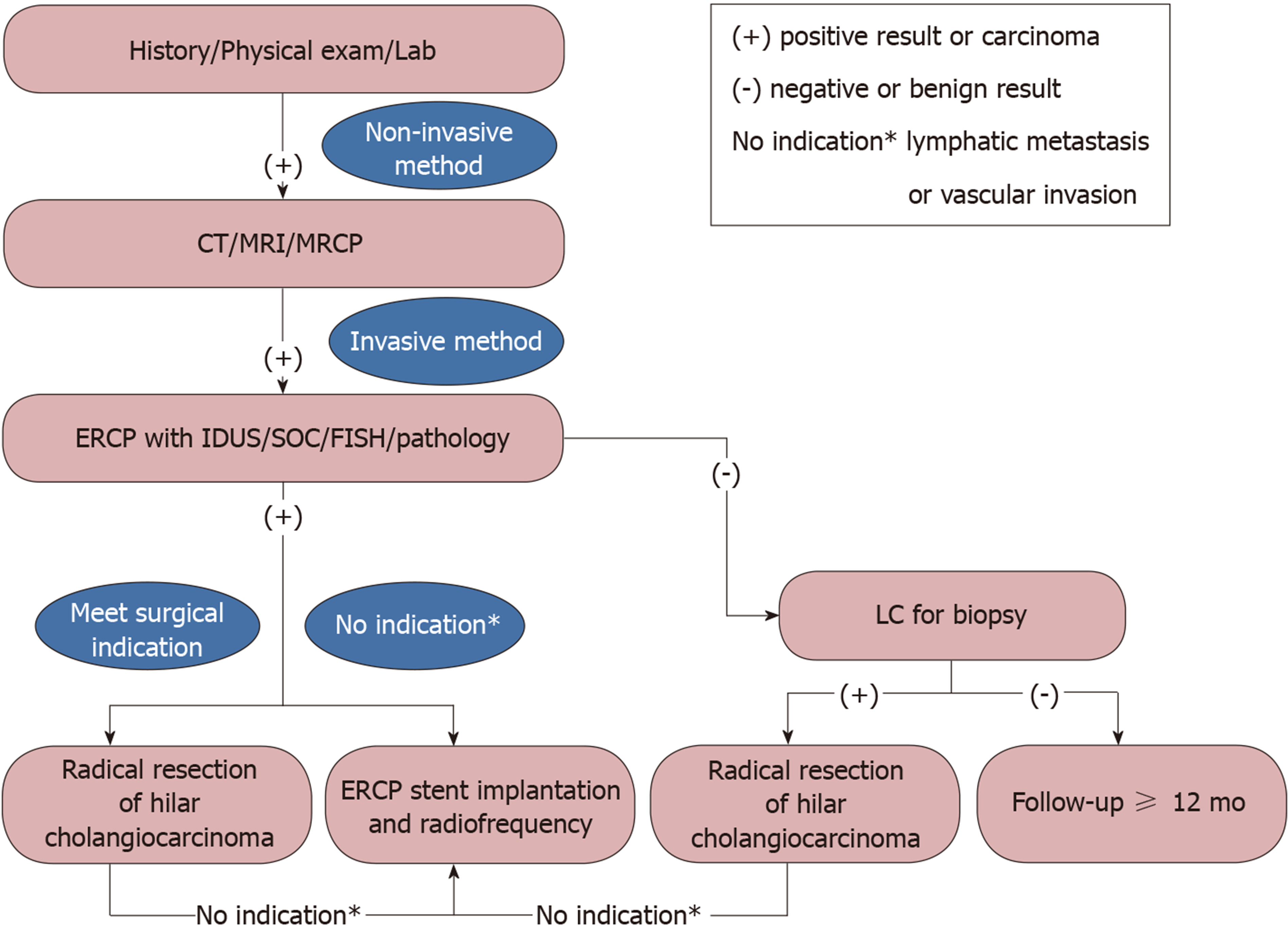
Figure 6 Various endoscopic and radiological imaging modalities for evaluation and preoperative diagnosis of gallbladder occupying lesions with bile duct invasion.
ERCP: Endoscopic retrograde cholangio-pancreatography.
- Citation: Wen LJ, Chen JH, Chen YJ, Liu K. Utility of multiple endoscopic techniques in differential diagnosis of gallbladder adenomyomatosis from gallbladder malignancy with bile duct invasion: A case report. World J Clin Cases 2020; 8(2): 464-470
- URL: https://www.wjgnet.com/2307-8960/full/v8/i2/464.htm
- DOI: https://dx.doi.org/10.12998/wjcc.v8.i2.464


