Copyright
©The Author(s) 2020.
World J Clin Cases. Jan 26, 2020; 8(2): 353-361
Published online Jan 26, 2020. doi: 10.12998/wjcc.v8.i2.353
Published online Jan 26, 2020. doi: 10.12998/wjcc.v8.i2.353
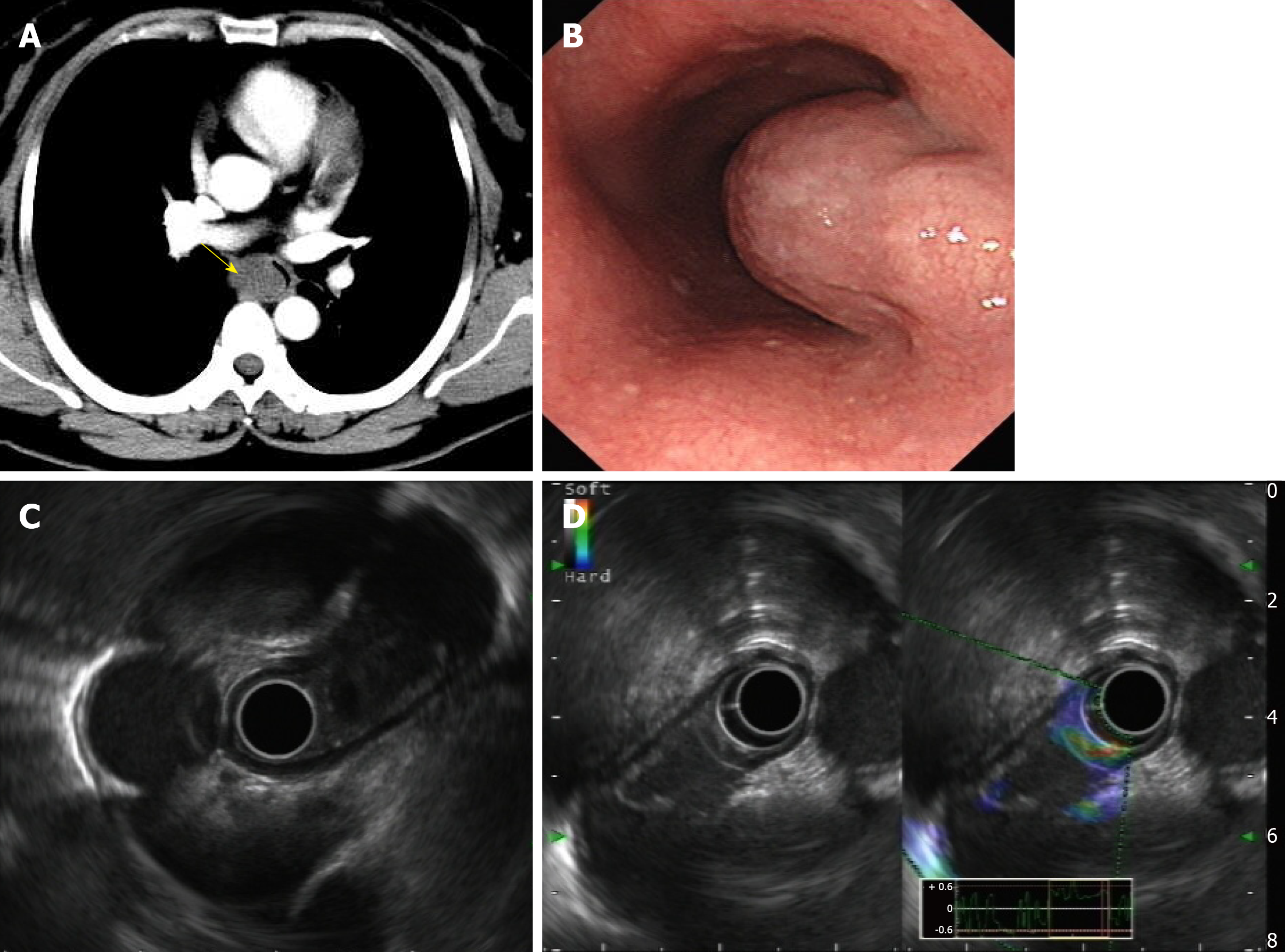
Figure 1 Enhanced thoracic computer tomography and endoscopic ultrasonography.
A: A slightly oval-shaped low-density lesion with clear boundary in the upper middle part of the esophagus in the enhanced thoracic computer tomography (yellow arrow); B: A submucosal mass was observed about 28 cm from the incisor with a gourd-like appearance; C: Probe EUS view; D: Contrast-enhanced ultrasonography showed slight enhancement around the lesion but no internal enhancement.
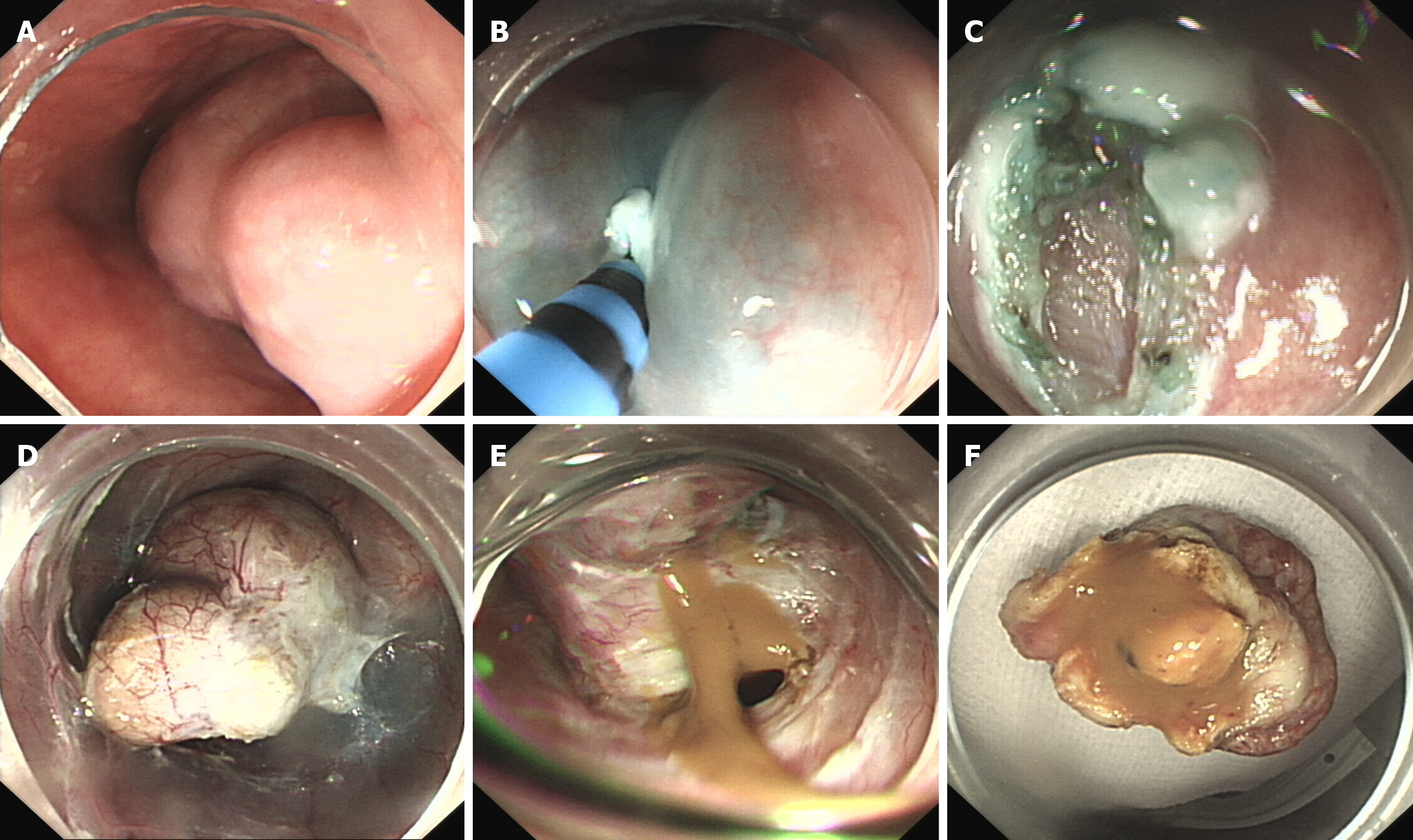
Figure 2 Endoscopic submucosal tunnel dissection of the esophageal bronchogenic cyst.
A: A submucosal mass was observed; B: Submucosal injection of normal saline with adrenaline and indigo carmine solution; C: HybridKnife was used to precut 2 cm of the esophageal mucosa; D: HybridKnife was used to perform submucosal separation, and the tumor was completely exposed; E: Yellow gelatinous liquid flowing out due to a small defect in the basilar cyst; F: The mass has been removed.
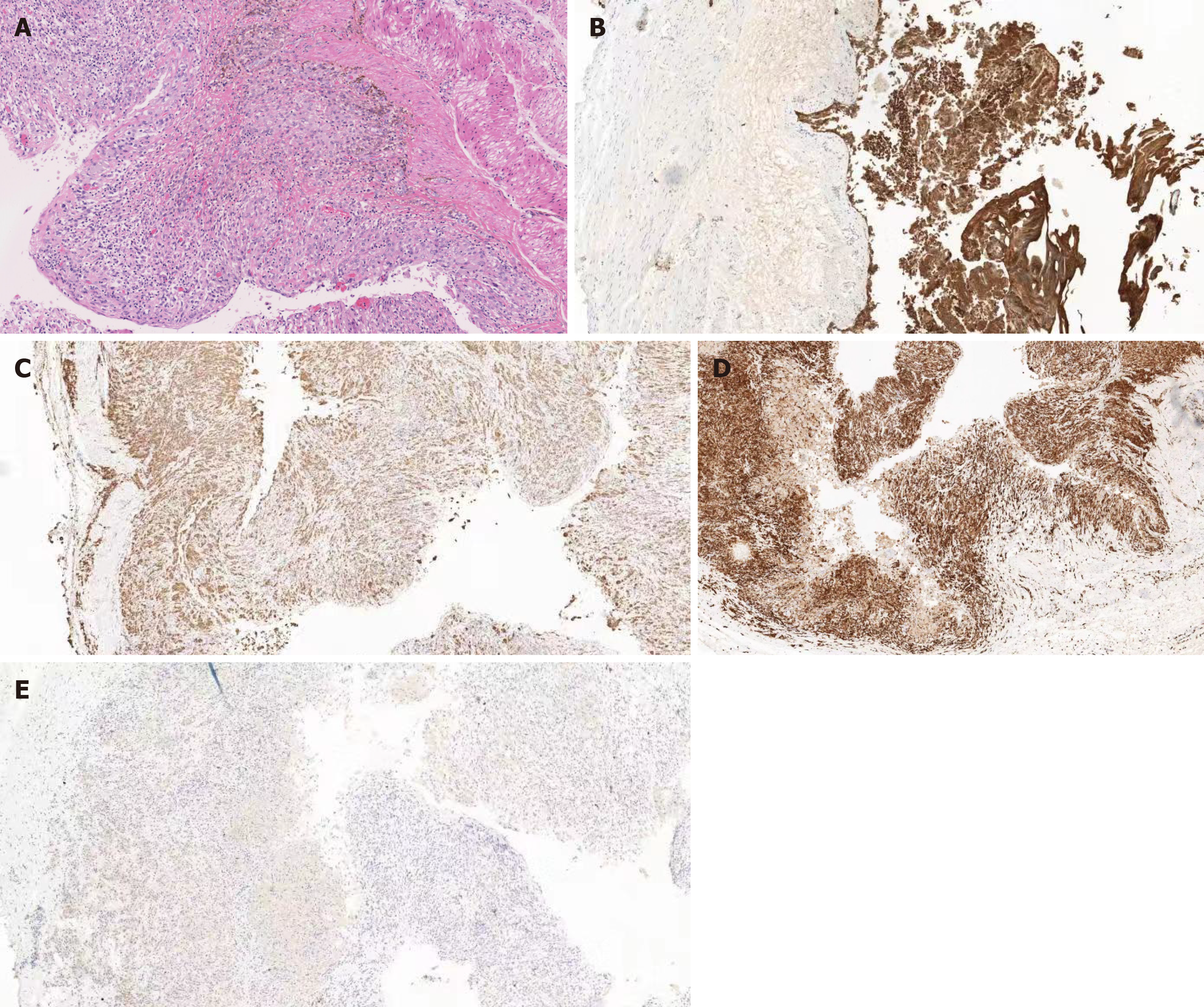
Figure 3 Histological examinations showed the specimen was consistent with bronchogenic cyst with obvious hyperplasia of histiocytes, and no dysplasia/malignancy was found.
A: HE stains × 200. B: CK (pan) (epithelium +) × 200; C: CD68 (histocyte +) × 200; D: CD163 (histocyte +) × 200; E: S-100 (-) × 200.
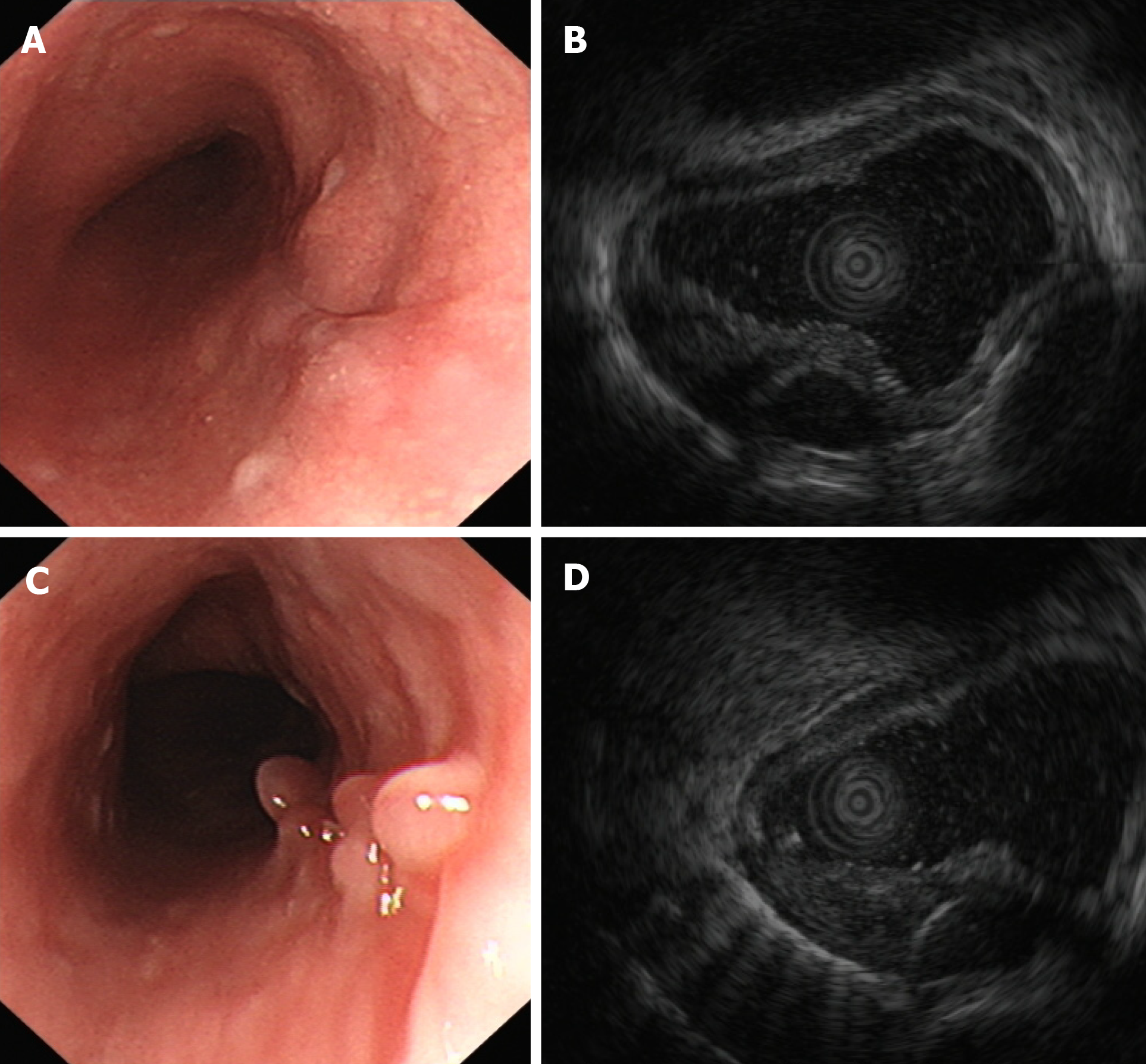
Figure 4 Endoscopic submucosal tunnel dissection and endoscopic ultrasonography.
A: Scar changes after endoscopic submucosal tunnel dissection; B: No cystic lesion was found within the original lesion under endoscopic ultrasonography; C: Granular mucosal uplift with smooth surface was observed; D: Local hypoechoic thickening of the intrinsic muscle layer was found.
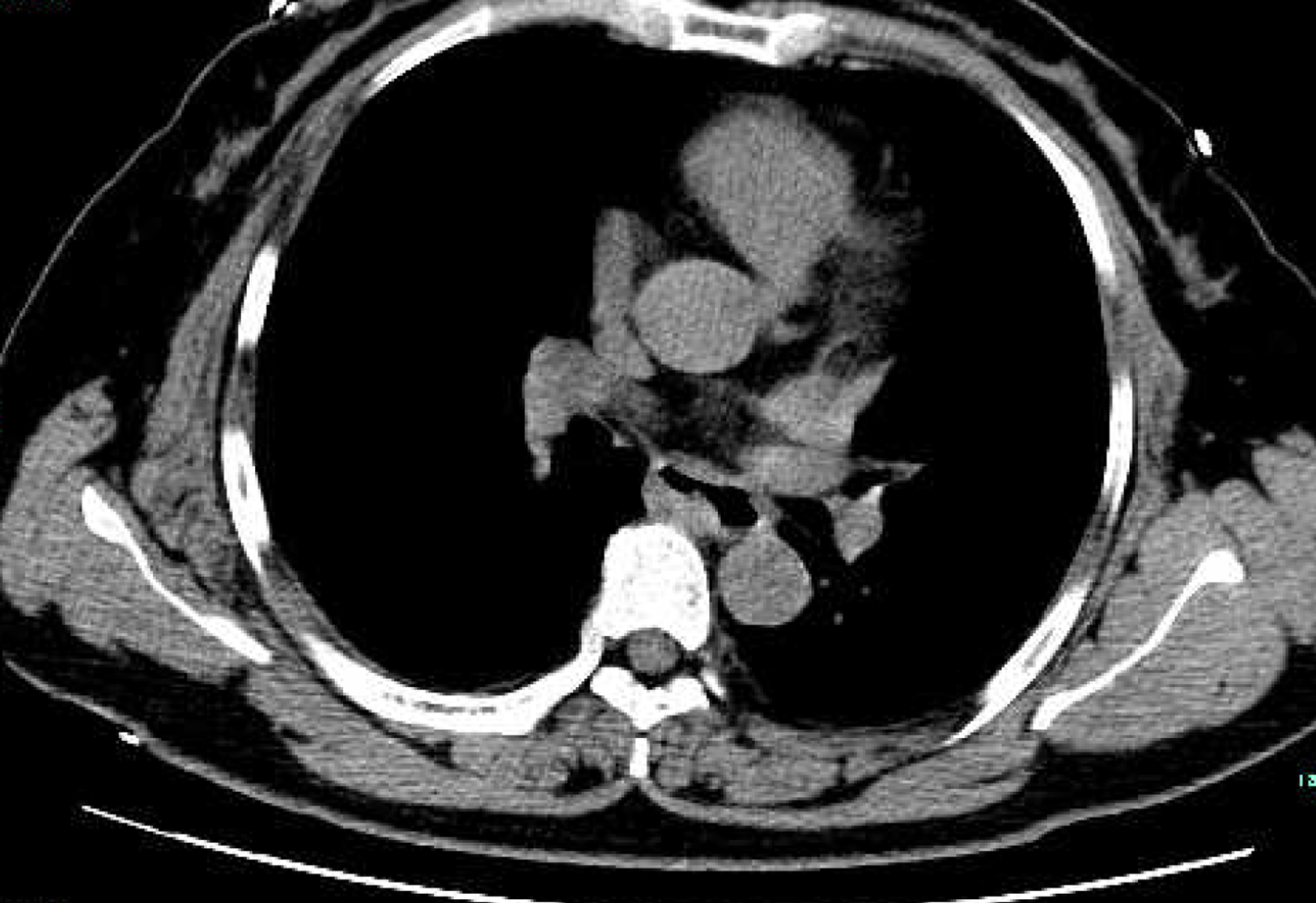
Figure 5 No obvious thickening of the esophagus wall, no abnormal high-density shadows in the esophagus lumen, no obvious expansion of the esophagus, and no significantly enlarged lymph node shadow was observed in the mediastinum.
- Citation: Zhang FM, Chen HT, Ning LG, Xu Y, Xu GQ. Esophageal bronchogenic cyst excised by endoscopic submucosal tunnel dissection: A case report. World J Clin Cases 2020; 8(2): 353-361
- URL: https://www.wjgnet.com/2307-8960/full/v8/i2/353.htm
- DOI: https://dx.doi.org/10.12998/wjcc.v8.i2.353


