Copyright
©The Author(s) 2020.
World J Clin Cases. Jan 26, 2020; 8(2): 306-312
Published online Jan 26, 2020. doi: 10.12998/wjcc.v8.i2.306
Published online Jan 26, 2020. doi: 10.12998/wjcc.v8.i2.306
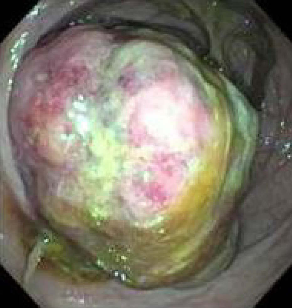
Figure 1 Findings at colonoscopy.
A large, friable, lobulated mass was identified in the mid-ascending colon.
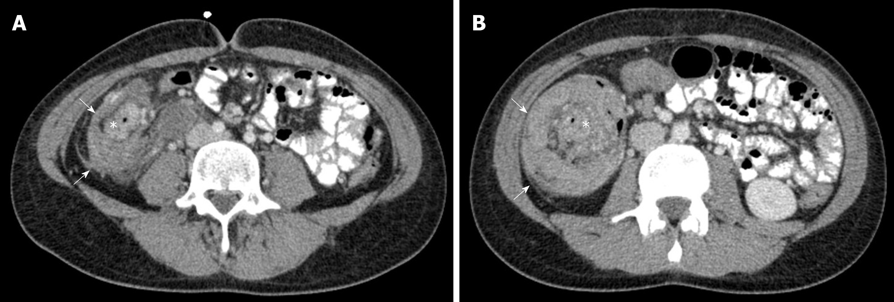
Figure 2 Abdominal and pelvic computed tomography scan.
A, B: The computed tomography scan showed an ileocolic intussusception (arrows) with an intraluminal mass lesion (*) acting as a lead point in the right colon.
- Citation: Giroux P, Collier A, Nowicki M. Recurrent lymphoma presenting as painless, chronic intussusception: A case report. World J Clin Cases 2020; 8(2): 306-312
- URL: https://www.wjgnet.com/2307-8960/full/v8/i2/306.htm
- DOI: https://dx.doi.org/10.12998/wjcc.v8.i2.306


