Copyright
©The Author(s) 2020.
World J Clin Cases. Sep 6, 2020; 8(17): 3841-3846
Published online Sep 6, 2020. doi: 10.12998/wjcc.v8.i17.3841
Published online Sep 6, 2020. doi: 10.12998/wjcc.v8.i17.3841
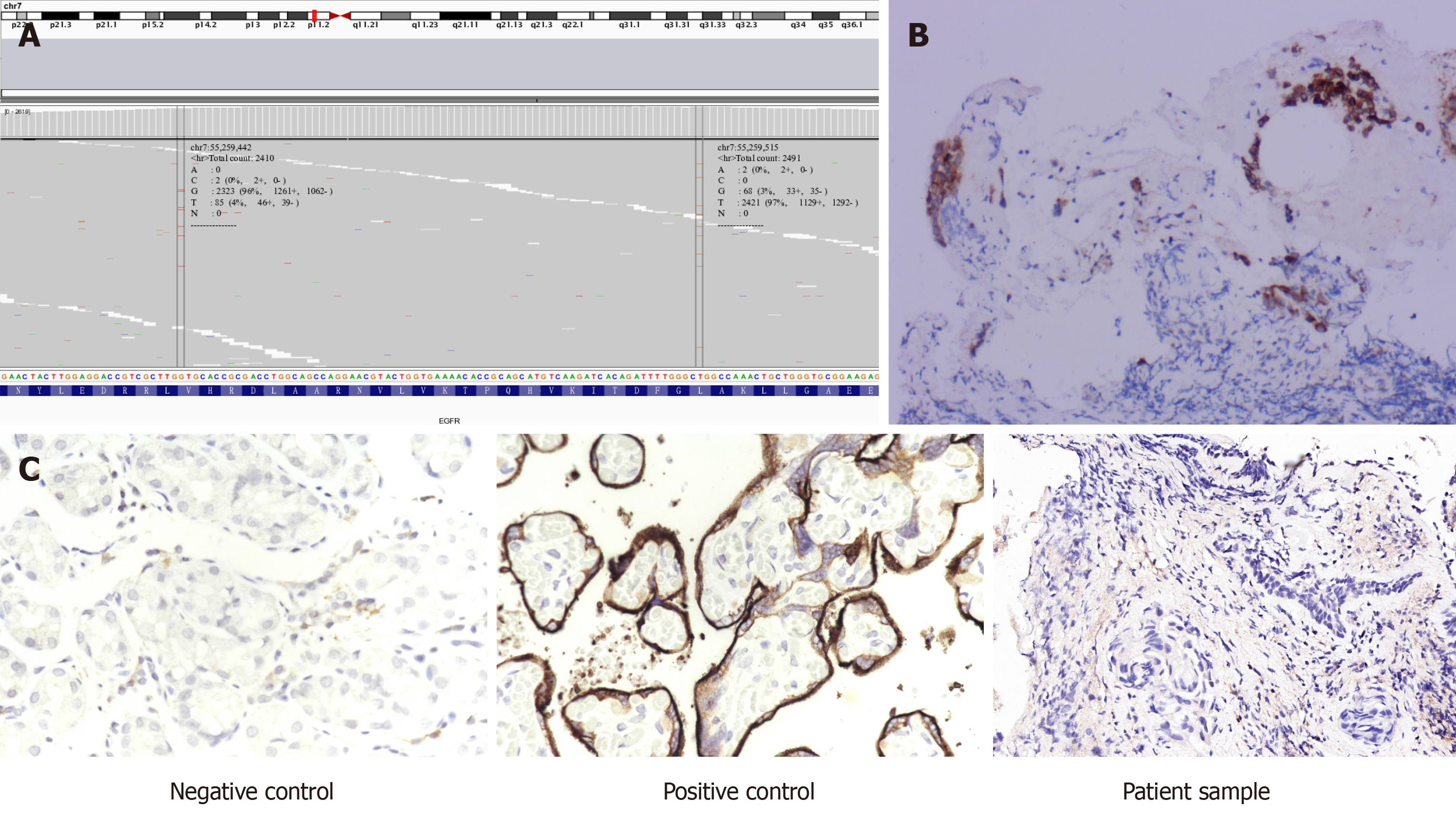
Figure 1 Next generation sequencing result and immunoassay of epidermal growth factor receptor and programmed death-ligand 1.
A: Next generation sequencing result of the patient’s tissue sample, showing variation in epidermal growth factor receptor exon 21; B: Epidermal growth factor receptor immunoassay; C: Programmed death-ligand 1 immunoassay.
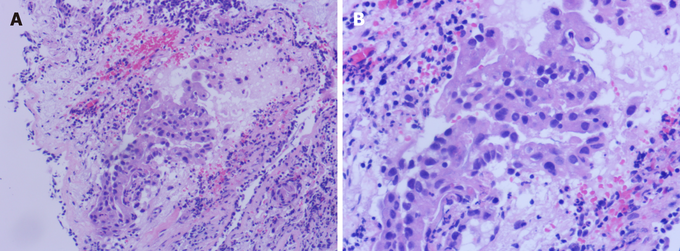
Figure 2 Representative images of hematoxylin and eosin-stained lung adenocarcinoma.
A and B: The tumor cells were characterized by the variation in nucleus size and shape, the deep staining and the increased nucleoplasm index, which was marked by a closed curve; Also, hematoxylin and eosin staining showed that the tumor cells invaded the surrounding tissue (A: 100 × and B: 200 ×).
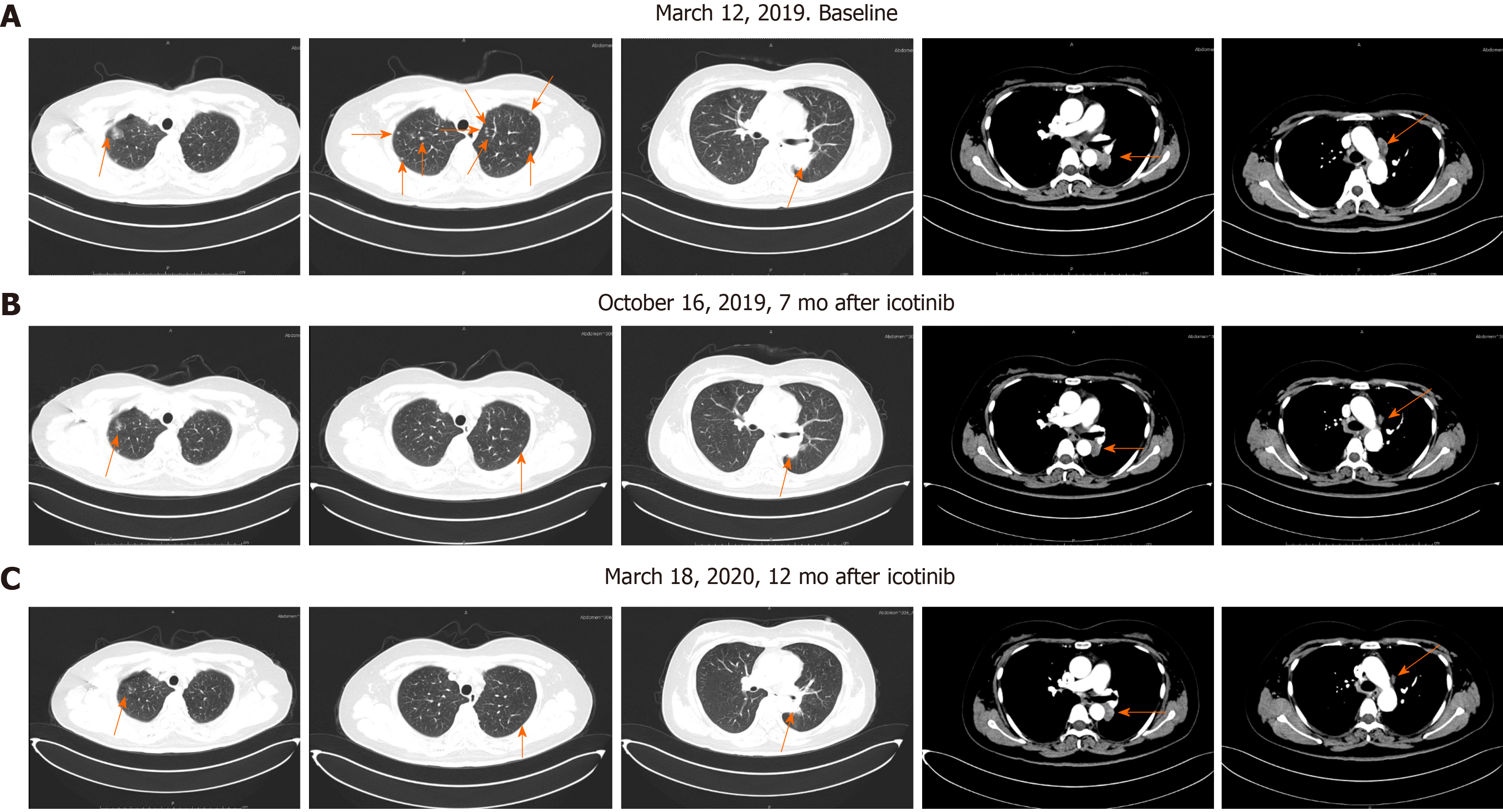
Figure 3 Primary lung cancer and mediastinal metastases before and after icotinib therapy.
A: Panel 1: Ground-glass nodules in the right upper lung; Panel 2: Multiple small nodules in both lungs; Panel 3: A mass shadow in the left lower lung dorsal segment; Panel 4: Soft tissue mass shadow in the left lower hilum of lung; Panel 5: Multiple swollen lymph nodes in the mediastinum, with largest located beside the aortic arch; B and C: Multiple small pulmonary nodules gradually reduced and disappeared, mediastinal lymph node metastasis decreased and periclavicular lymph node metastasis decreased and disappeared.
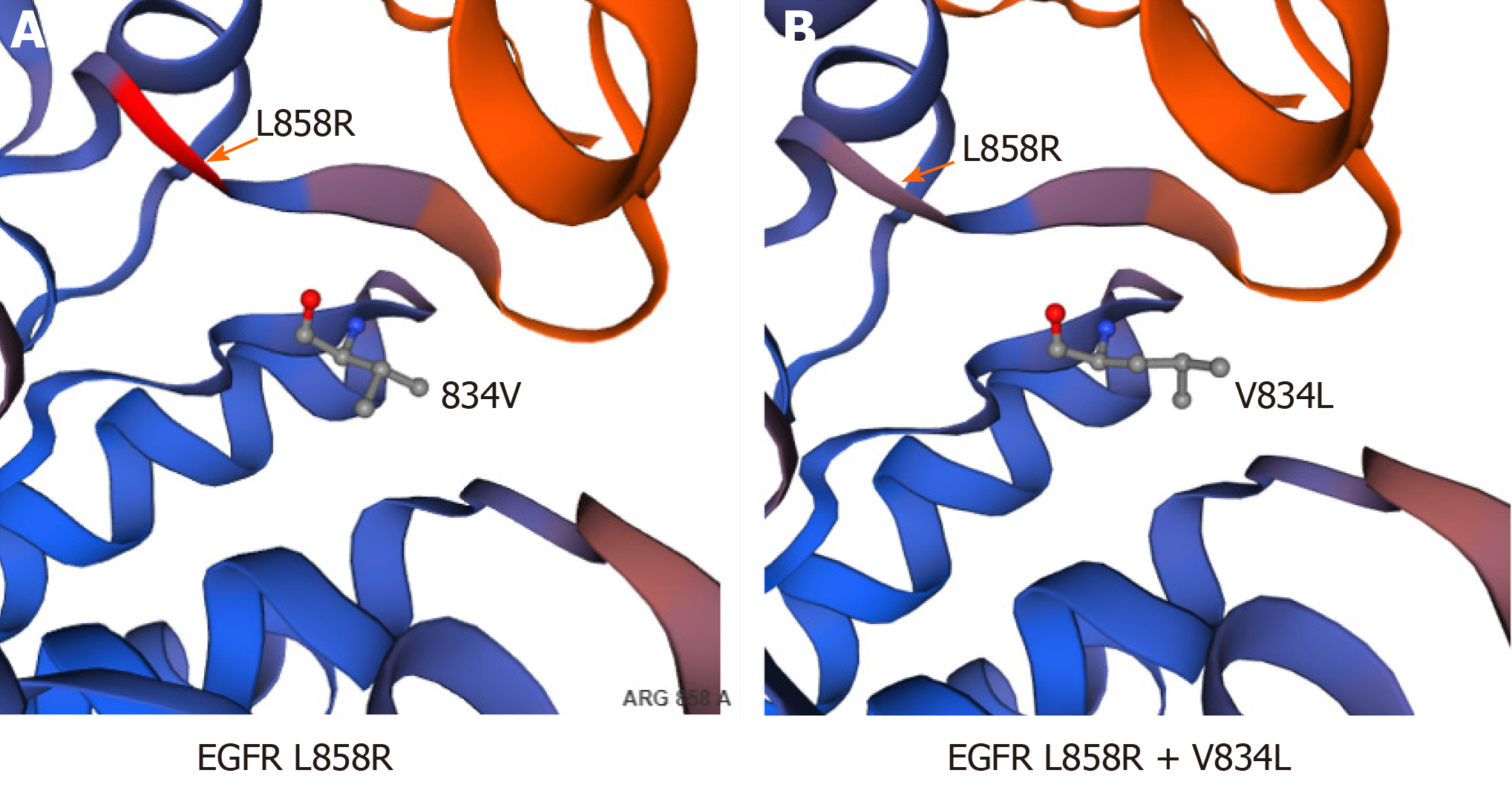
Figure 4 Structural analysis of epidermal growth factor receptor L858R and V834L mutants.
- Citation: Zhai SS, Yu H, Gu TT, Li YX, Lei Y, Zhang HY, Zhen TH, Gao YG. Lung adenocarcinoma harboring rare epidermal growth factor receptor L858R and V834L mutations treated with icotinib: A case report. World J Clin Cases 2020; 8(17): 3841-3846
- URL: https://www.wjgnet.com/2307-8960/full/v8/i17/3841.htm
- DOI: https://dx.doi.org/10.12998/wjcc.v8.i17.3841


