Copyright
©The Author(s) 2020.
World J Clin Cases. Aug 6, 2020; 8(15): 3334-3340
Published online Aug 6, 2020. doi: 10.12998/wjcc.v8.i15.3334
Published online Aug 6, 2020. doi: 10.12998/wjcc.v8.i15.3334
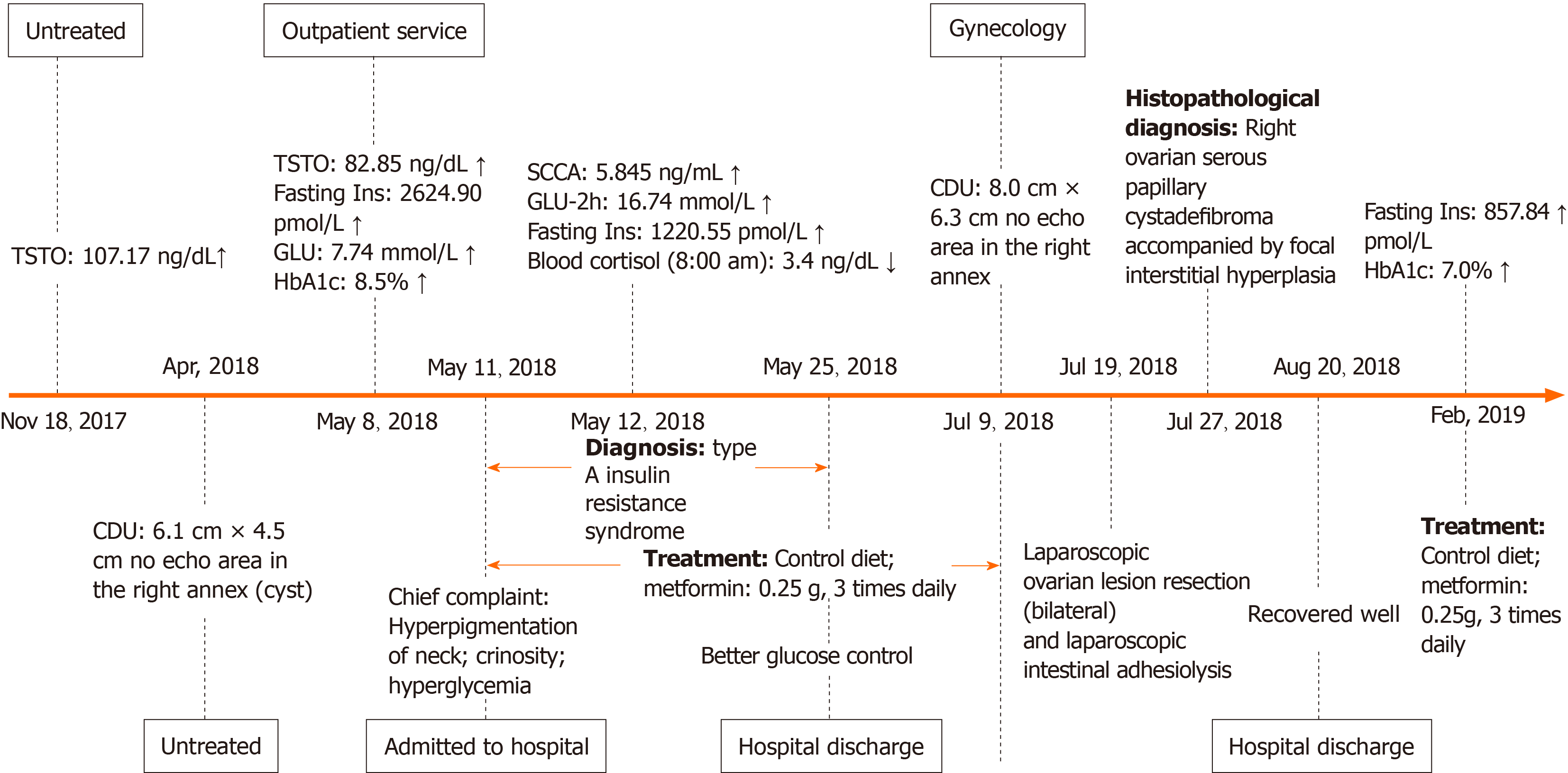
Figure 1 Clinical symptoms and laboratory indices in the 14-year-old girl patient.
“↑” indicates above the normal range; “↓” indicates blow the normal range. TSTO: Testosterone; CDU: Color Doppler ultrasound; Ins: Insulin; GLU: Glucose; HbA1c: Hemoglobin A1c; SCCA: Squamous cell carcinoma antigen.
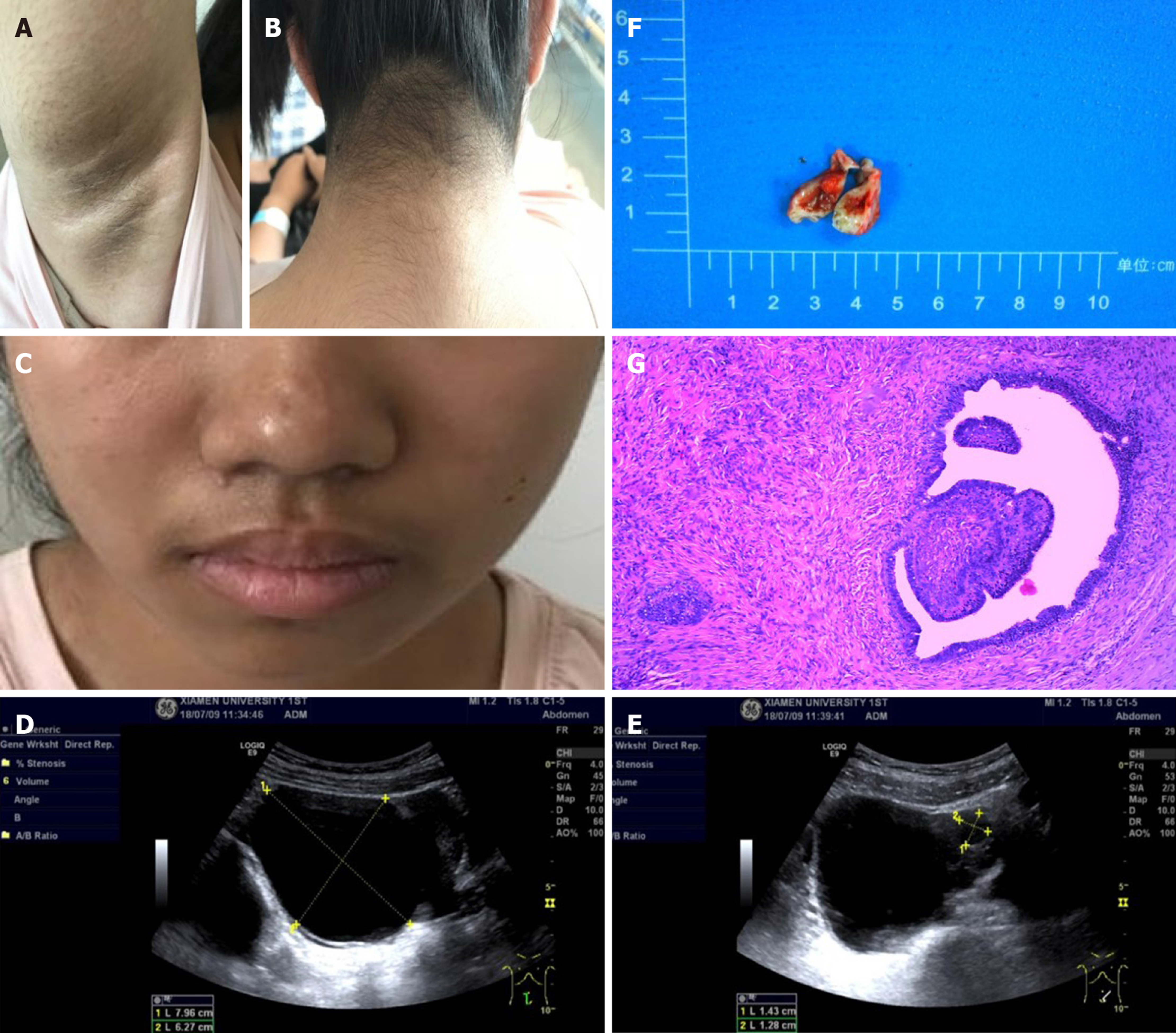
Figure 2 Clinical characteristics, genetic diagnosis, and histopathological diagnosis in this patient.
A: Axillary skin is hairy, dark, coarse, and warty, with small papillae; B: The skin on the neck is black, thickened, rough, and warty, with small papillae; C: Whiskers are present on the upper lip; D: A cystic lesion was found at the upper right side of the uterus, approximately 8.0 cm × 6.3 cm, with well-defined borders; E: The cystic lesion included a larger quasi-circular anechoic area, approximately 1.4 cm × 1.2 cm; F: The nodular mass of the right ovary is cystic and solid in section, with a pale brown solid area approximately 1.5 cm × 1.2 cm × 0.8 cm in size; G: Right ovarian serous papillary cystadenofibroma accompanied by focal interstitial hyperplasia (hematoxylin-eosin, × 40).
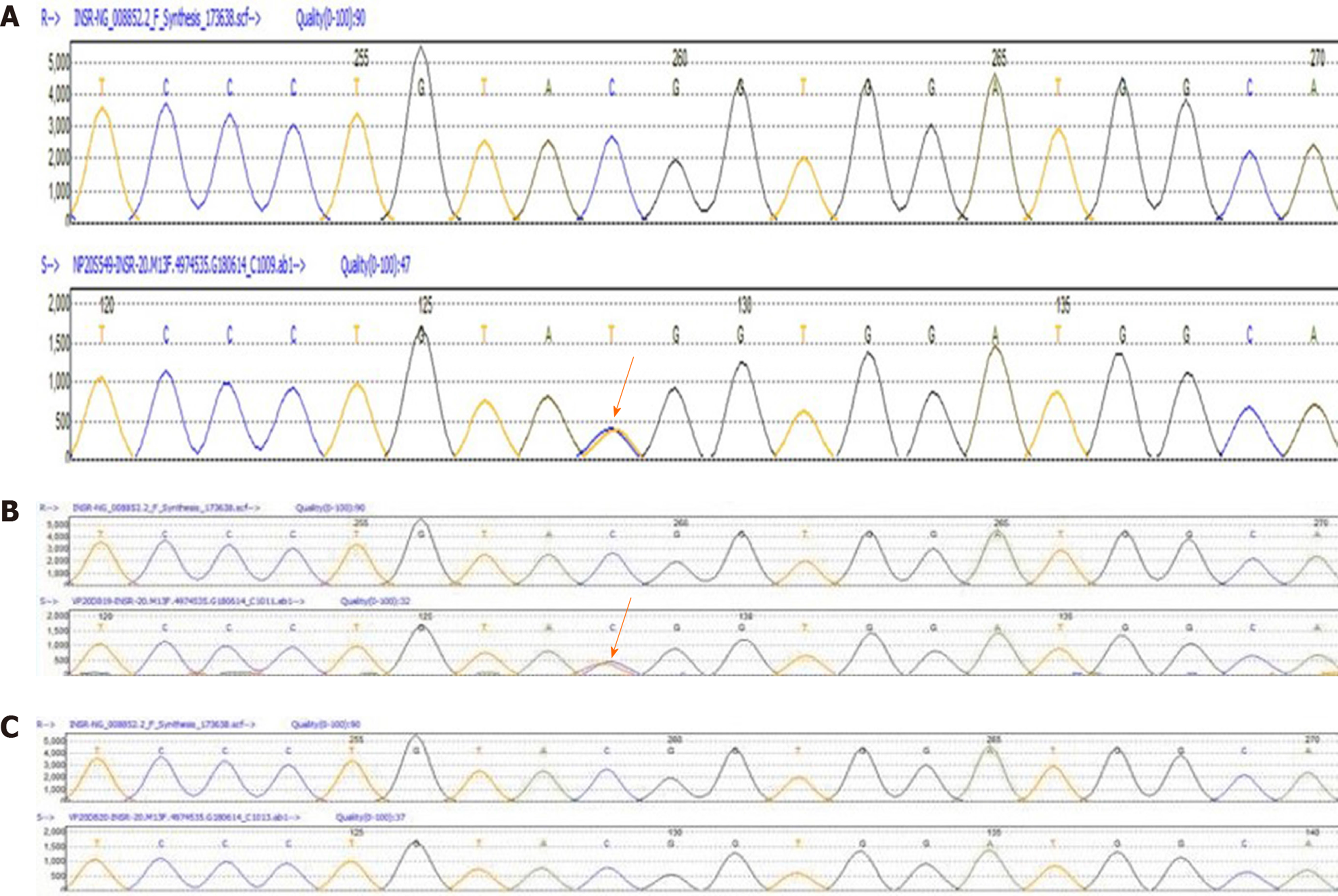
Figure 3 Heterozygous mutation of the insulin receptor was observed in the patient and her father, as detected by Sanger sequencing (A-C).
A missense variant exists in exon 20 (c.3601C>T [p.Arg1201Trp]).
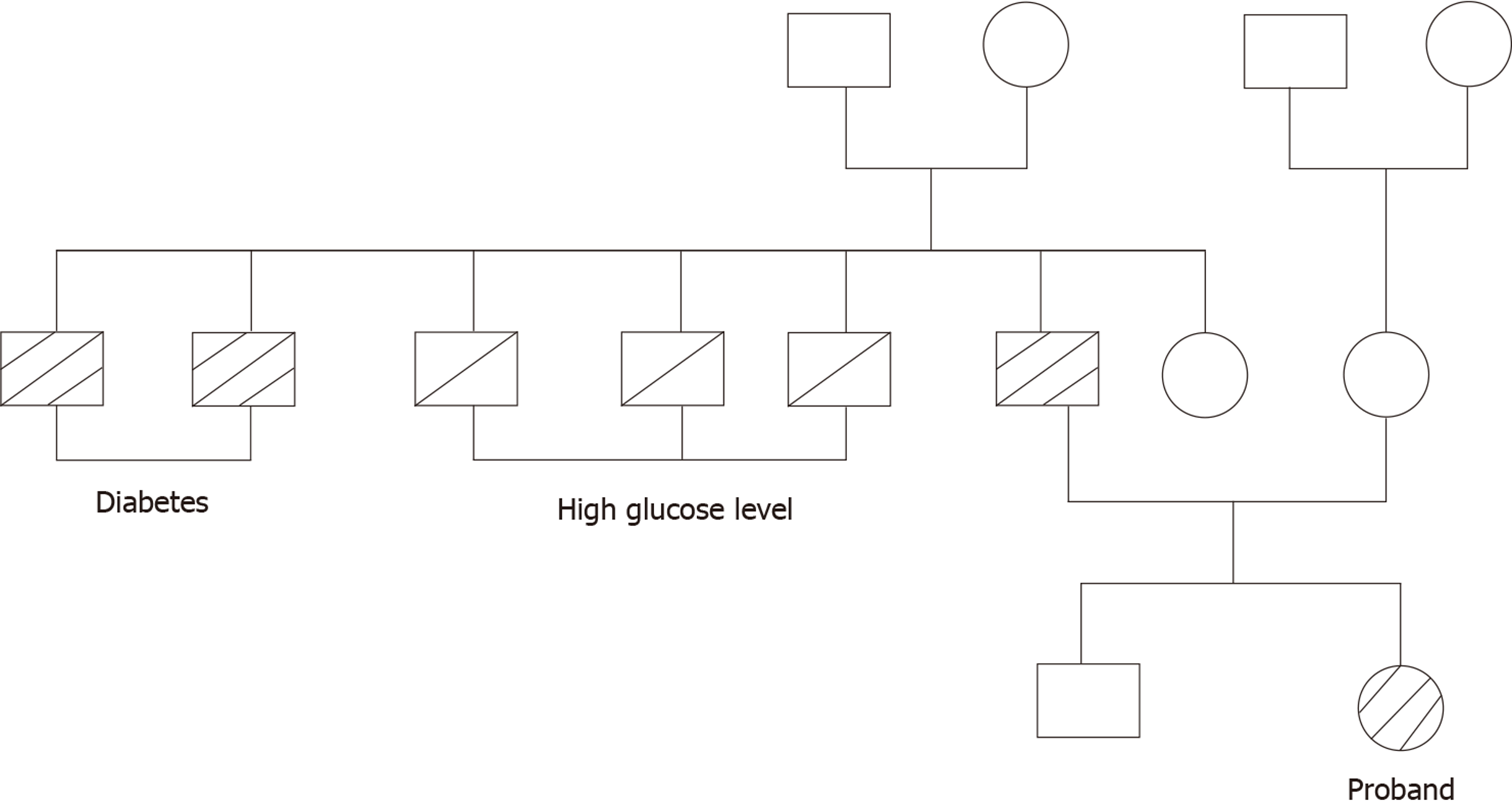
Figure 4 Family history of diabetes.
- Citation: Yan FF, Huang BK, Chen YL, Zhuang YZ, You XY, Liu CQ, Li XJ. Coexistence of ovarian serous papillary cystadenofibroma and type A insulin resistance syndrome in a 14-year-old girl: A case report. World J Clin Cases 2020; 8(15): 3334-3340
- URL: https://www.wjgnet.com/2307-8960/full/v8/i15/3334.htm
- DOI: https://dx.doi.org/10.12998/wjcc.v8.i15.3334


