Copyright
©The Author(s) 2020.
World J Clin Cases. Aug 6, 2020; 8(15): 3314-3319
Published online Aug 6, 2020. doi: 10.12998/wjcc.v8.i15.3314
Published online Aug 6, 2020. doi: 10.12998/wjcc.v8.i15.3314
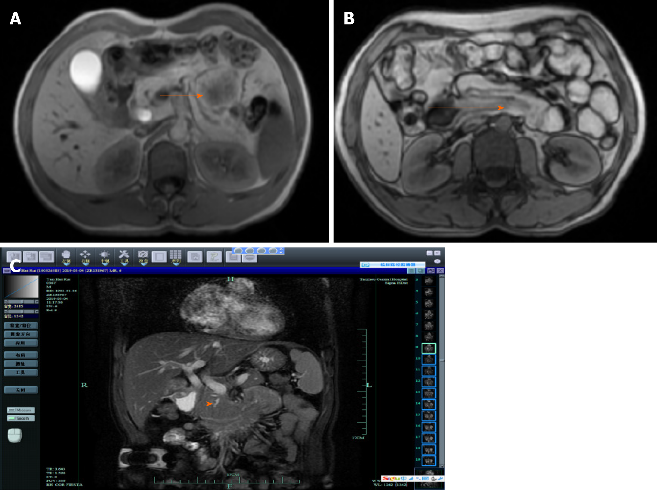
Figure 1 Magnetic resonance cholangiopancreatography.
A: Magnetic resonance cholangiopancreatography (MRCP) revealed an abnormal mass signal in the distal duodenum; B: MRCP revealed duodeno-duodenal intussusception; C: Location of mass and intussusception after MRCP recombination.
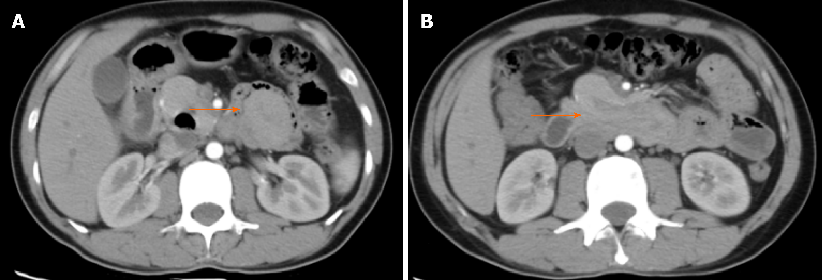
Figure 2 Computed tomography.
A: Computed tomography (CT) revealed an abnormal mass signal in the distal duodenum; B: CT revealed duodeno-duodenal intussusception.
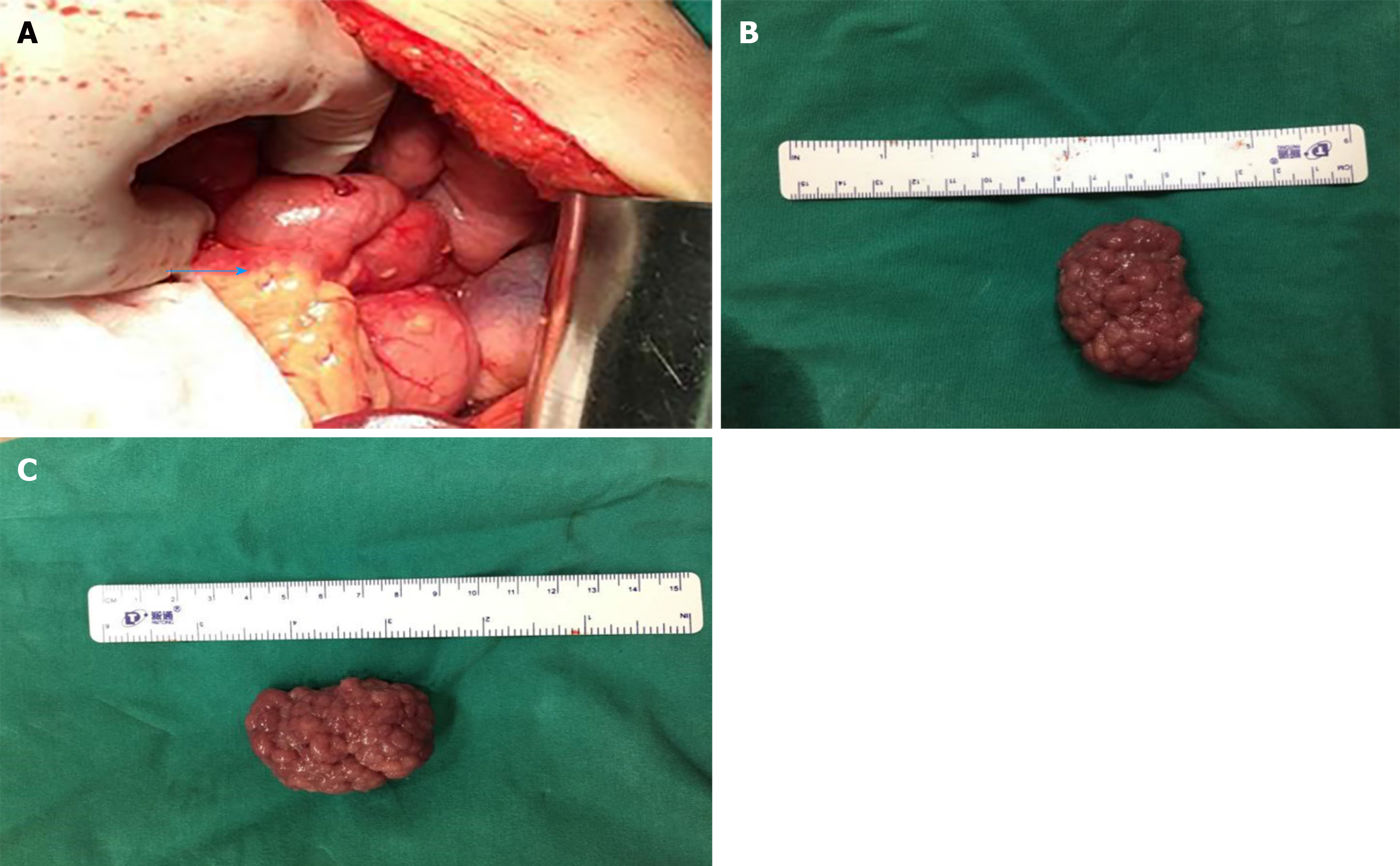
Figure 3 Ascending part of the duodenum and diameter of the adenoma.
A: Intussusception of the descending part of the duodenum entered the horizontal part and combined closely; B: Diameter of the longitudinal axis of the adenoma was approximately 3.5 cm; C: Diameter of the transverse axis of the adenoma was approximately 5.0 cm.
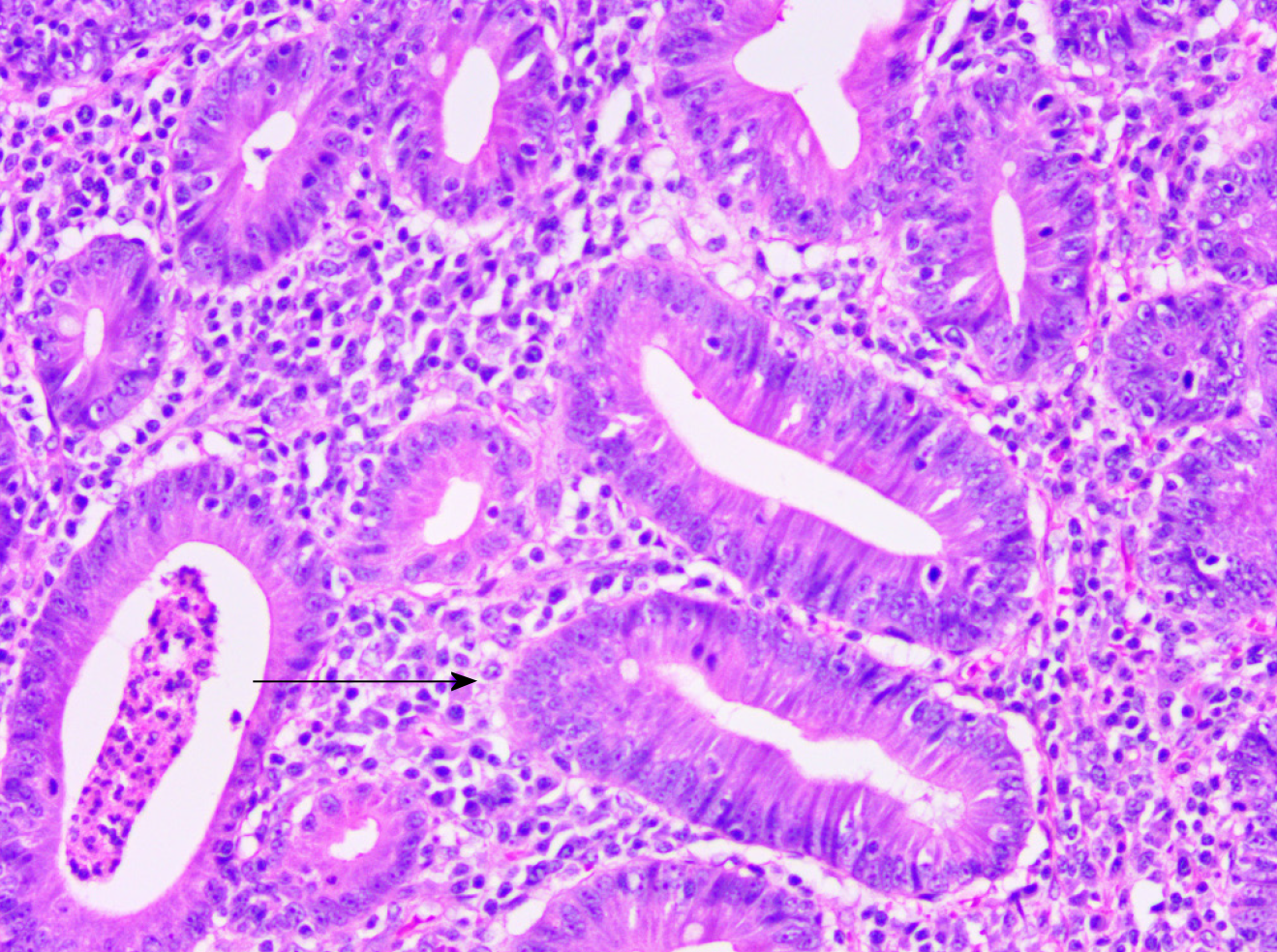
Figure 4 Postoperative routine pathology revealed tubular-villous adenoma with low-grade glandular intraepithelial neoplasia (local high-grade intraepithelial neoplasia).
- Citation: Wang KP, Jiang H, Kong C, Wang LZ, Wang GY, Mo JG, Jin C. Adult duodenal intussusception with horizontal adenoma: A rare case report. World J Clin Cases 2020; 8(15): 3314-3319
- URL: https://www.wjgnet.com/2307-8960/full/v8/i15/3314.htm
- DOI: https://dx.doi.org/10.12998/wjcc.v8.i15.3314


