Copyright
©The Author(s) 2020.
World J Clin Cases. Aug 6, 2020; 8(15): 3177-3187
Published online Aug 6, 2020. doi: 10.12998/wjcc.v8.i15.3177
Published online Aug 6, 2020. doi: 10.12998/wjcc.v8.i15.3177
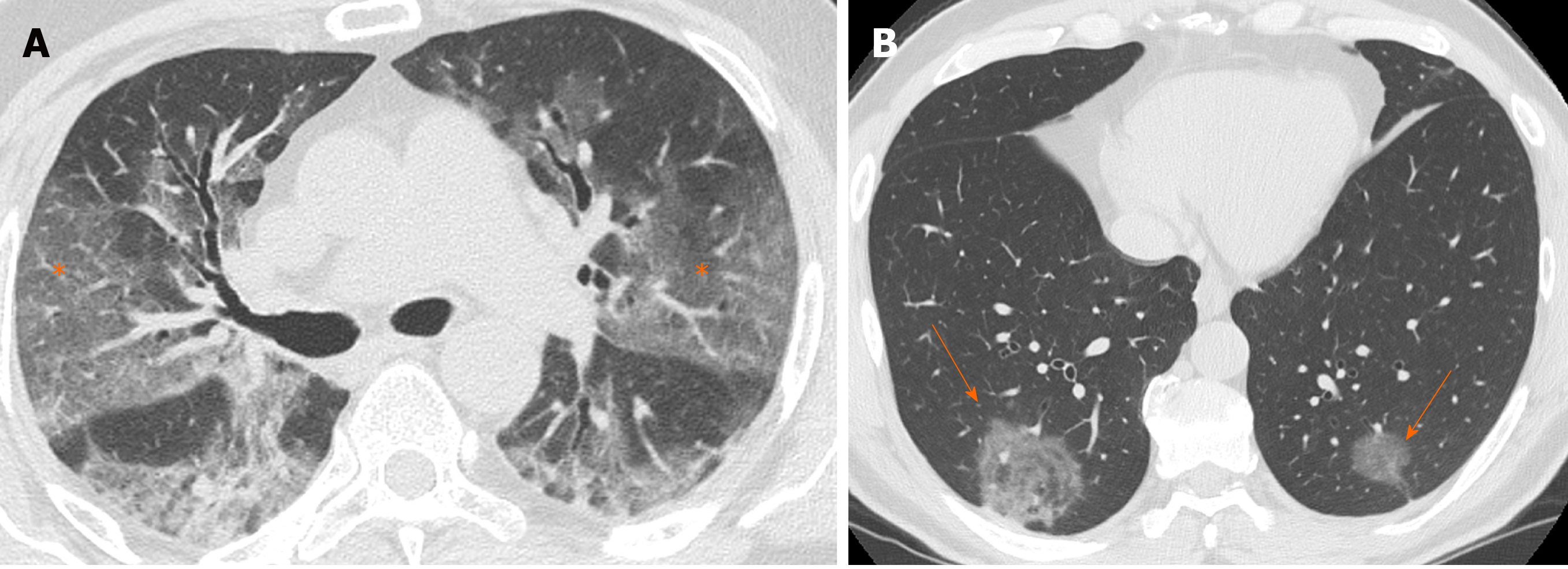
Figure 1 Computed tomography images of ground glass opacity.
A: 59-year man admitted to the emergency room presenting fever and cough. Chest computed tomography showed diffuse bilateral confluent and predominantly linear ground glass opacities (asterisks); and B: 60-year man admitted to the emergency room presenting cough and abdominal pain. Chest computed tomography showed bilateral round ground glass opacities (orange arrows).
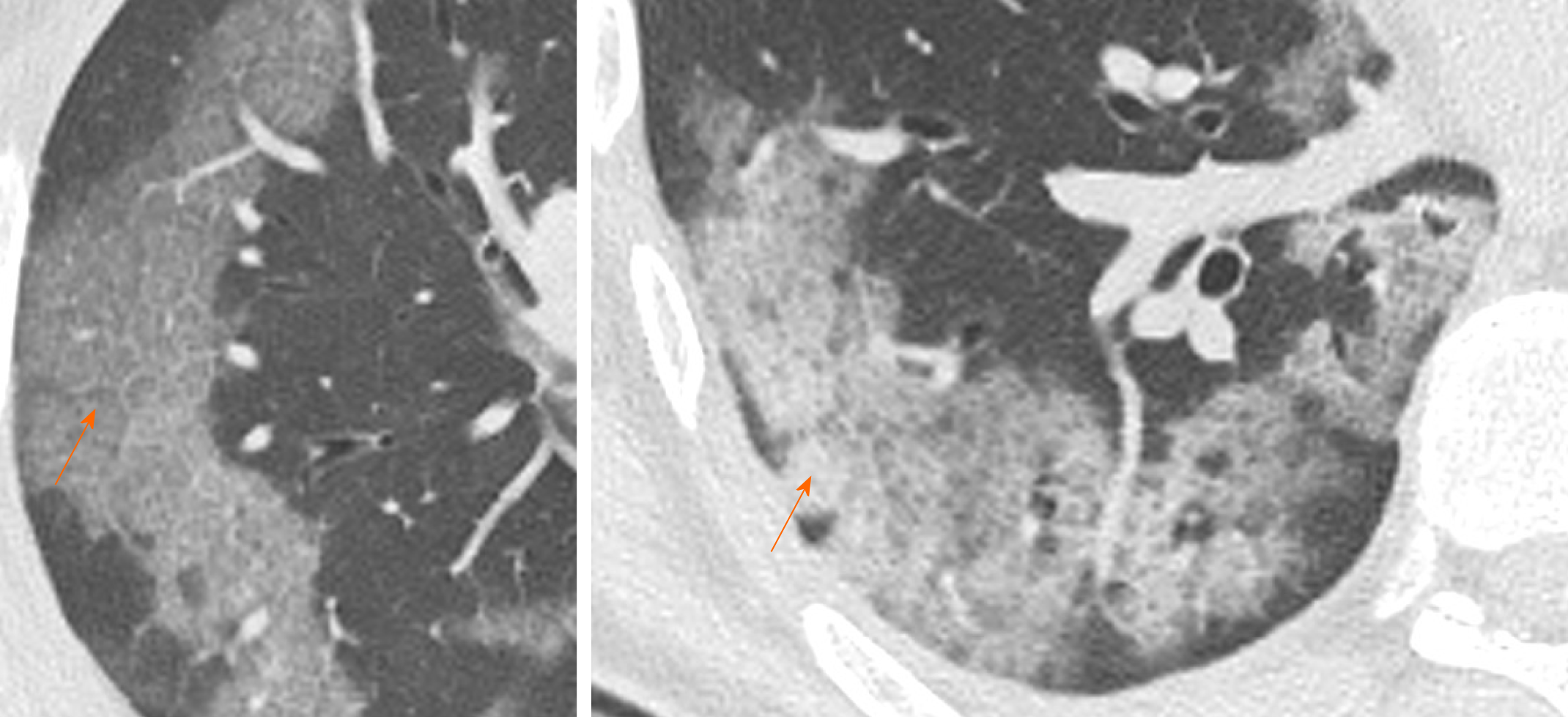
Figure 2 Computed tomography Images of crazy paving.
70-year and 55-year men admitted to the emergency room presenting fever, cough and worsening dyspnea. Chest computed tomography showed confluent and predominantly patchy ground glass opacities with pronounced peripheral distribution and interlobular and intralobular septal thickening (crazy paving pattern), in right upper lobe and right lower lobe, respectively (orange arrows).
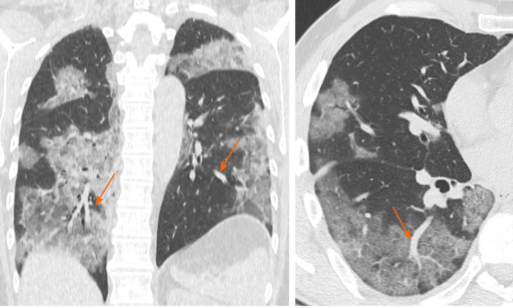
Figure 3 Computed tomography images of vessel enlargement.
57-year woman admitted to the emergency room presenting cough and fever. Chest computed tomography showed segmental and subsegmental vessels’ enlargement (orange arrows) and multi-lobe ground glass opacities parenchymal involvement.
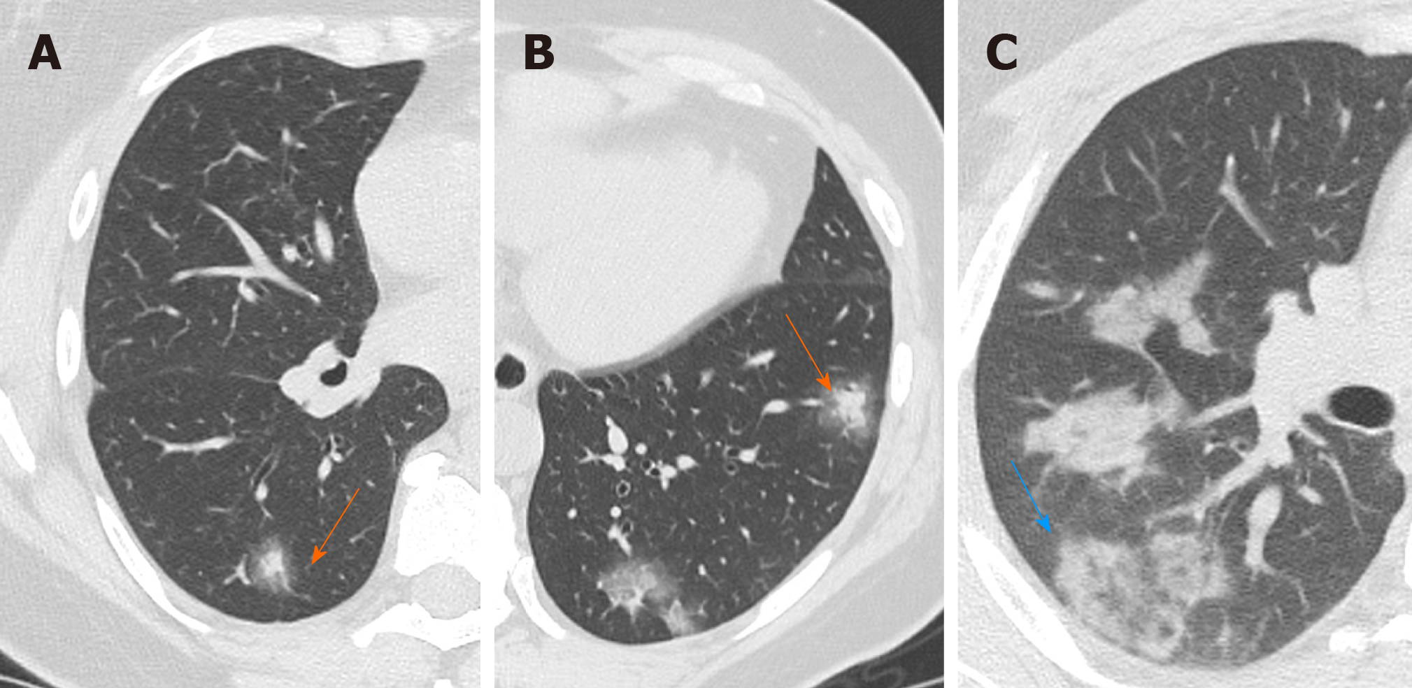
Figure 4 Computed tomography Images of halo sign-reversed halo sign.
A and B: 50-year woman admitted to the emergency room presenting fever and cough. Chest computed tomography showed nodular consolidations surrounded by a zone of ground glass attenuation in both lower lobes indicating the halo sign (orange arrows); and C: 36-year man admitted to the emergency room presenting fever and dyspnea. Chest computed tomography showed a focal rounded ground glass opacity surrounded by an incomplete ring-like consolidation indicating the reverse halo sign (blue arrow).
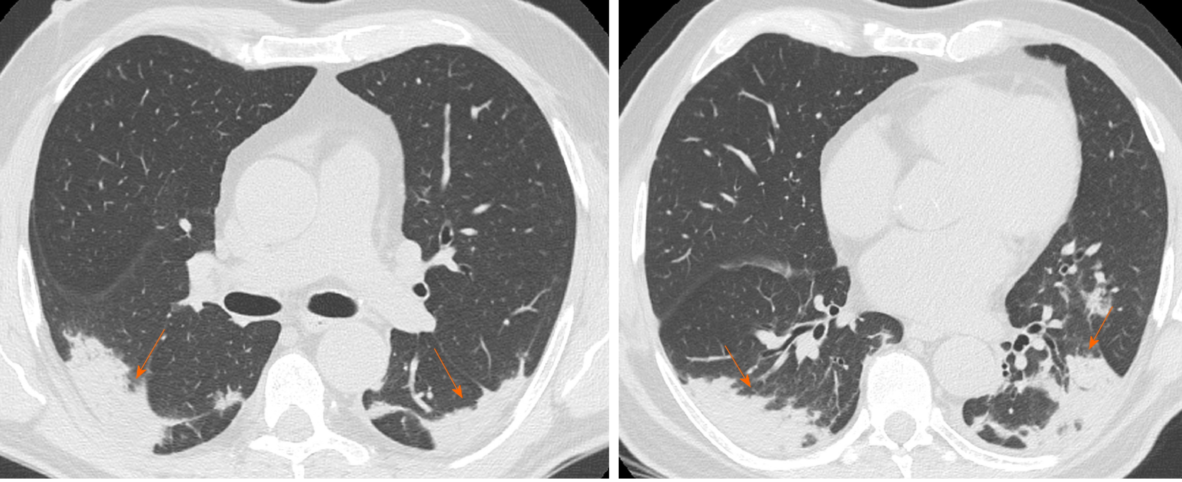
Figure 5 Computed tomography images of consolidation.
66-year man admitted to the emergency room with cough and fever. Chest computed tomography showed bilateral subpleural consolidations without air bronchogram, with posterior distribution in both upper and lower lobes (orange arrows).
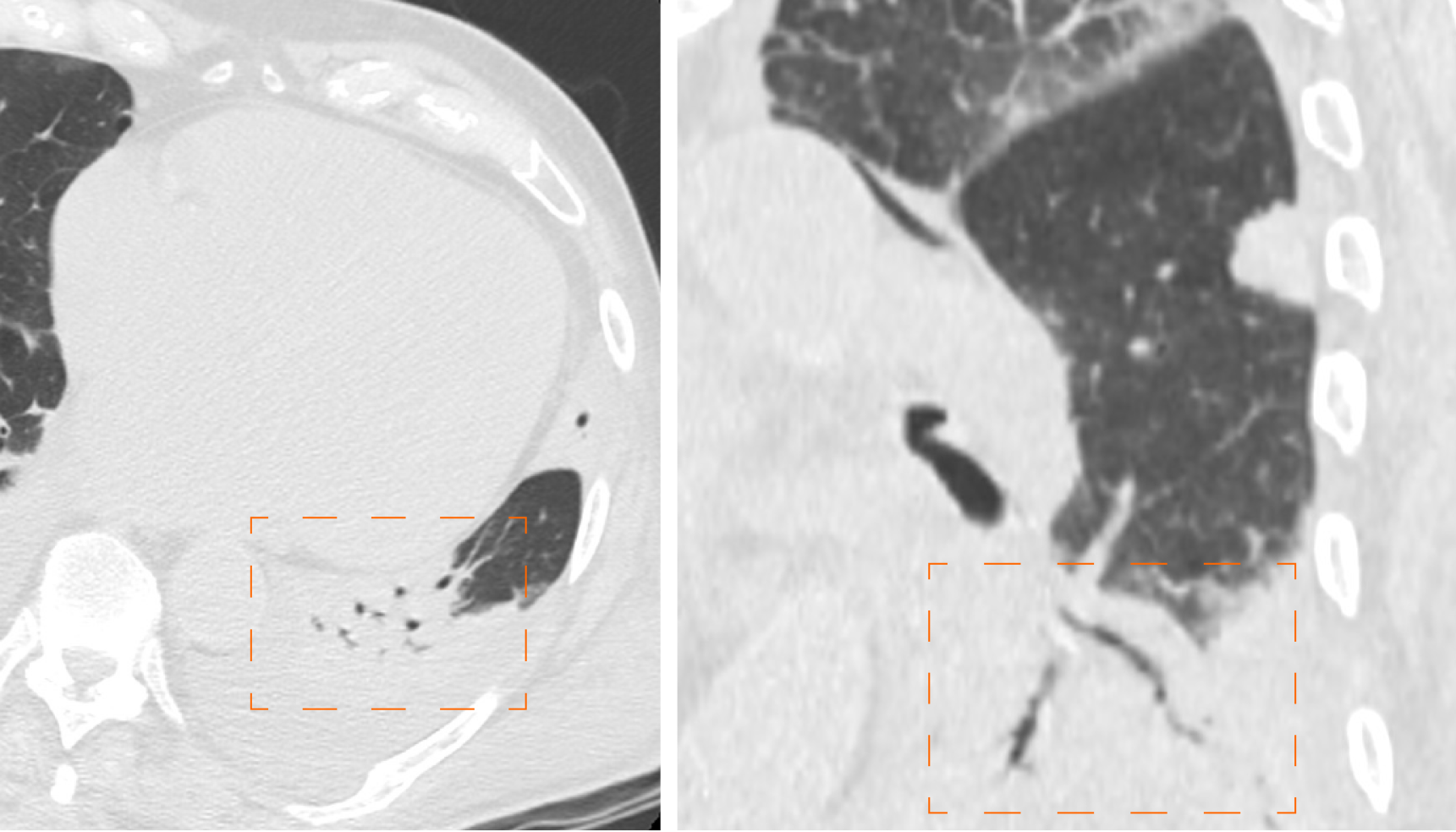
Figure 6 Computed tomography images of air bronchogram.
92-year man admitted to the emergency room presenting cough, fever and worsening dyspnea. Chest computed tomography showed pulmonary consolidation with air bronchogram in left lower lobe (dashed orange frames).
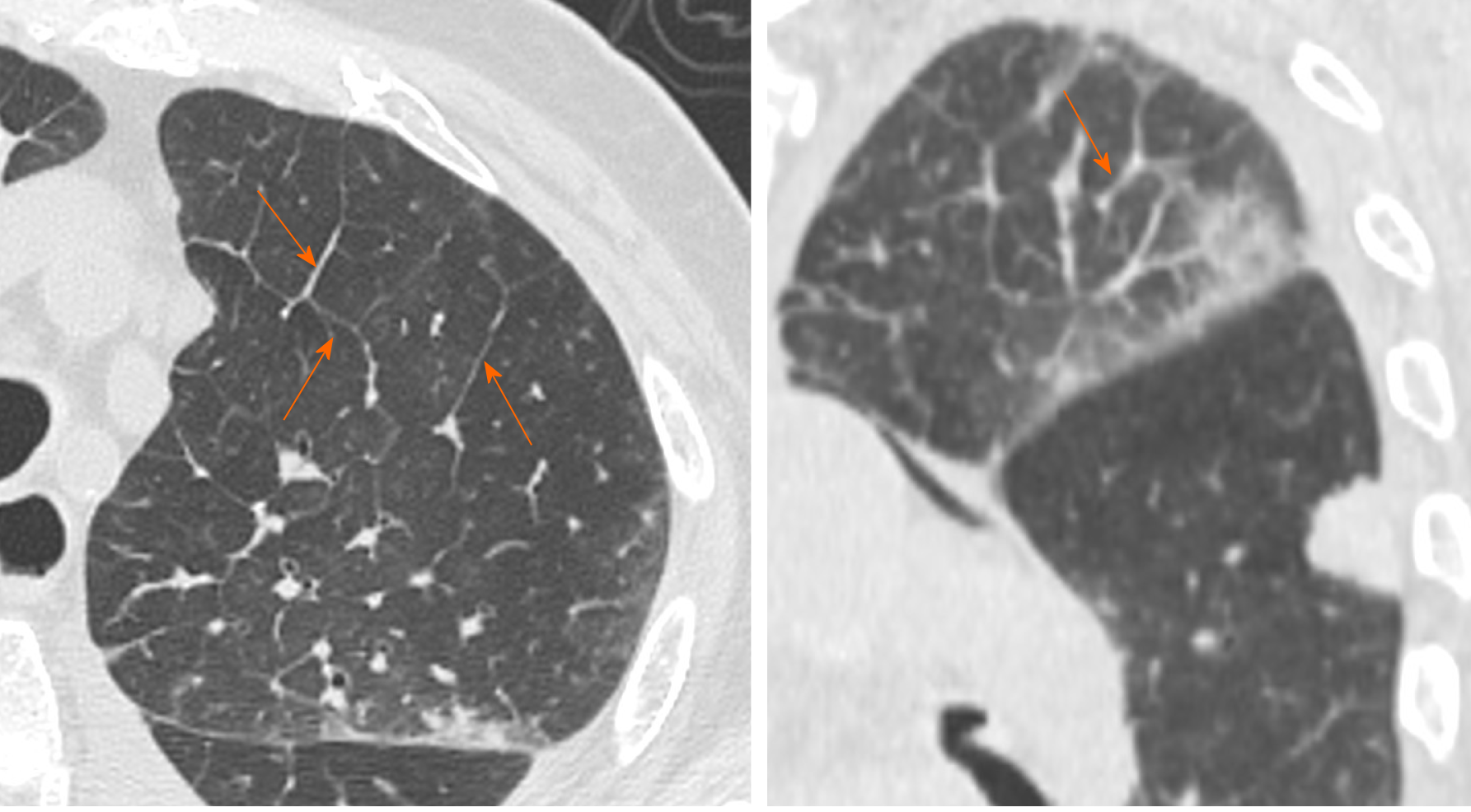
Figure 7 Computed tomography images of interstitial septal thickening.
92-year man admitted to the emergency room presenting cough, fever and worsening dyspnea. Chest computed tomography showed interlobular septal thickening predominantly in left upper lobe (orange arrows).
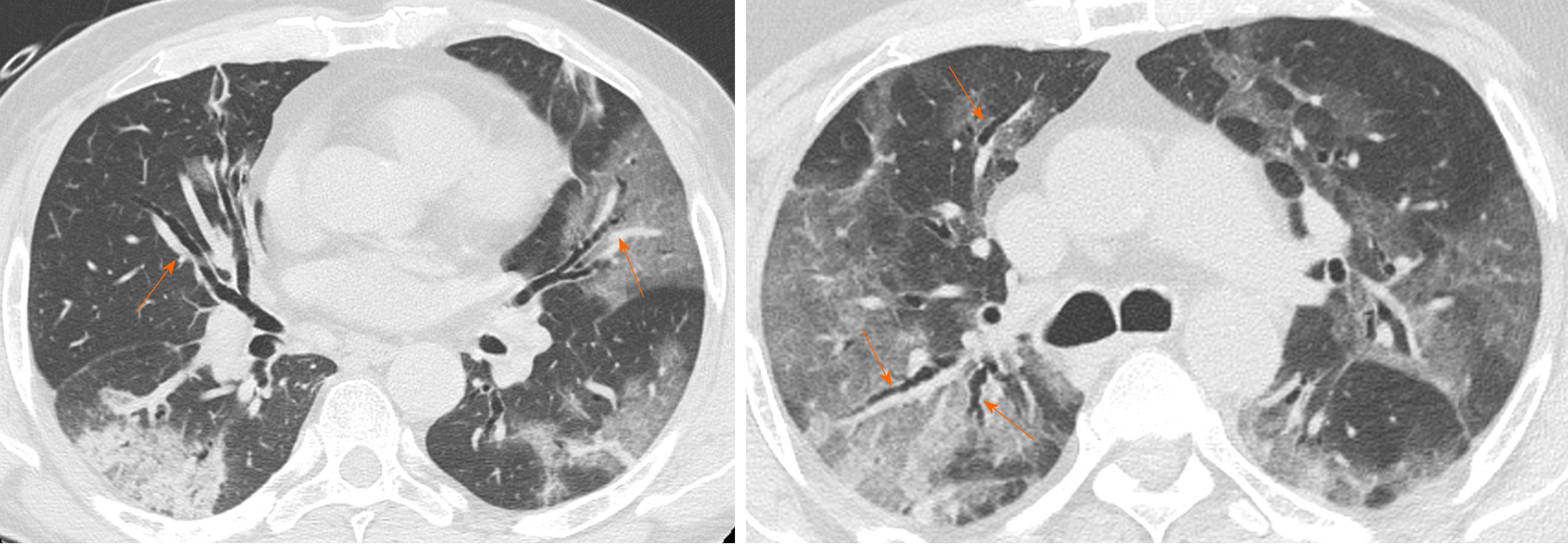
Figure 8 Computed tomography images of bronchiectasis.
65-year man admitted to the emergency room presenting fever and worsening dyspnea. Chest computed tomography showed diffuse bilateral bronchiectasis (orange arrows) and multi-lobar parenchymal involvement.
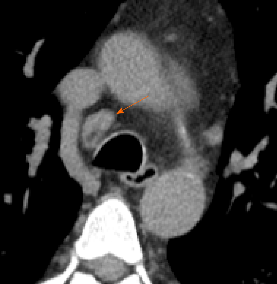
Figure 9 Computed tomography images of lymphadenopathy.
61-year man admitted to the emergency room presenting cough and dyspnea. Chest computed tomography showed a lymphadenopathy (short axis > 10 mm) in right lower paratracheal station (orange arrow).
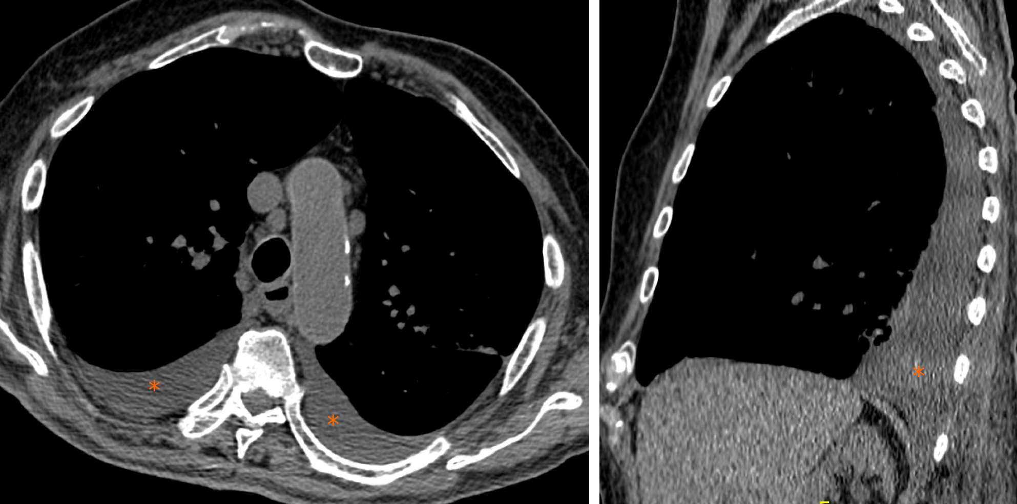
Figure 10 Computed tomography images of pleural effusion.
88-year man admitted to the emergency room presenting cough, fever and dyspnea. Chest computed tomography showed bilateral pleural effusion (asterisks).
- Citation: Caruso D, Polidori T, Guido G, Nicolai M, Bracci B, Cremona A, Zerunian M, Polici M, Pucciarelli F, Rucci C, Dominicis CD, Girolamo MD, Argento G, Sergi D, Laghi A. Typical and atypical COVID-19 computed tomography findings. World J Clin Cases 2020; 8(15): 3177-3187
- URL: https://www.wjgnet.com/2307-8960/full/v8/i15/3177.htm
- DOI: https://dx.doi.org/10.12998/wjcc.v8.i15.3177


