Copyright
©The Author(s) 2020.
World J Clin Cases. Jul 26, 2020; 8(14): 3130-3135
Published online Jul 26, 2020. doi: 10.12998/wjcc.v8.i14.3130
Published online Jul 26, 2020. doi: 10.12998/wjcc.v8.i14.3130
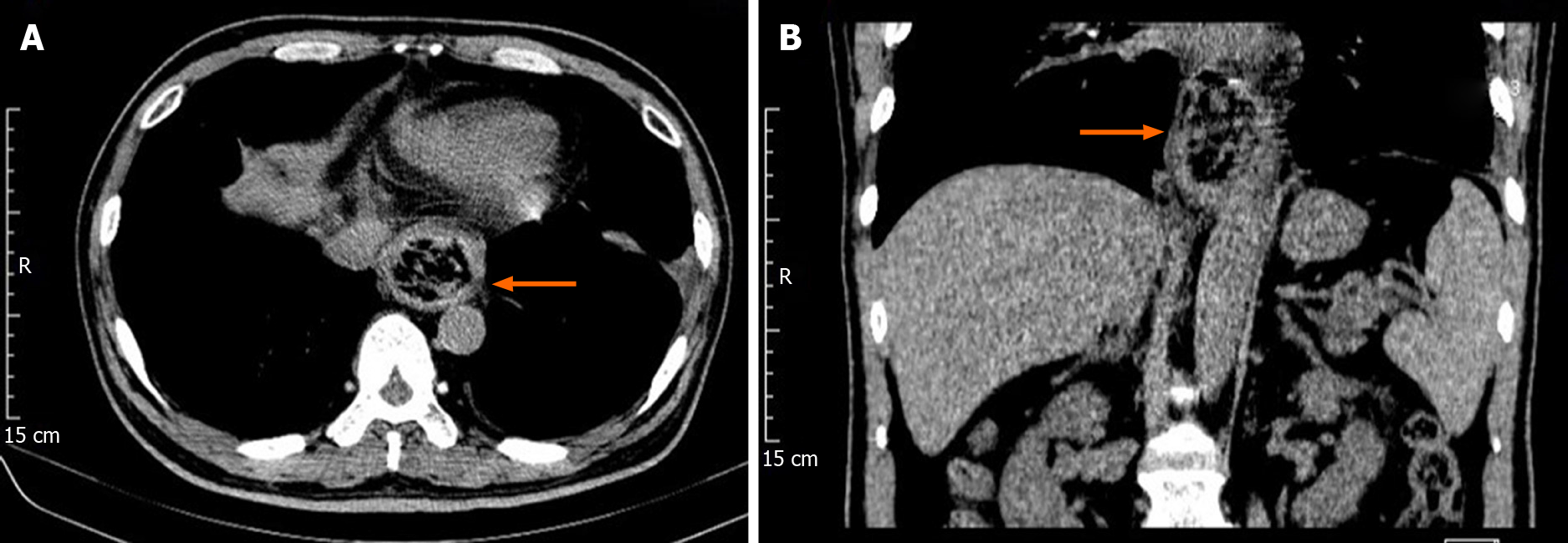
Figure 1 Computed tomography findings.
A: Coronal plane; B: Sagittal plane. Computed tomography scan revealed dilated lower esophagus, thickening of the esophageal wall, a mass-like lesion with flocculent high-density shadow and gas bubbles in the esophageal lumen (orange arrow).
Figure 2 Gastroscope images.
A: Large immovable brown bezoars embedded in the lower esophagus; B: Multiple mucosal edema, erosions and superficial ulcers were observed in the lower esophagus after endoscopic fragmentation; C: Bezoars removed from the esophagus.
- Citation: Zhang FH, Ding XP, Zhang JH, Miao LS, Bai LY, Ge HL, Zhou YN. Acute esophageal obstruction caused by reverse migration of gastric bezoars: A case report. World J Clin Cases 2020; 8(14): 3130-3135
- URL: https://www.wjgnet.com/2307-8960/full/v8/i14/3130.htm
- DOI: https://dx.doi.org/10.12998/wjcc.v8.i14.3130


