Copyright
©The Author(s) 2020.
World J Clin Cases. Jul 26, 2020; 8(14): 3114-3121
Published online Jul 26, 2020. doi: 10.12998/wjcc.v8.i14.3114
Published online Jul 26, 2020. doi: 10.12998/wjcc.v8.i14.3114
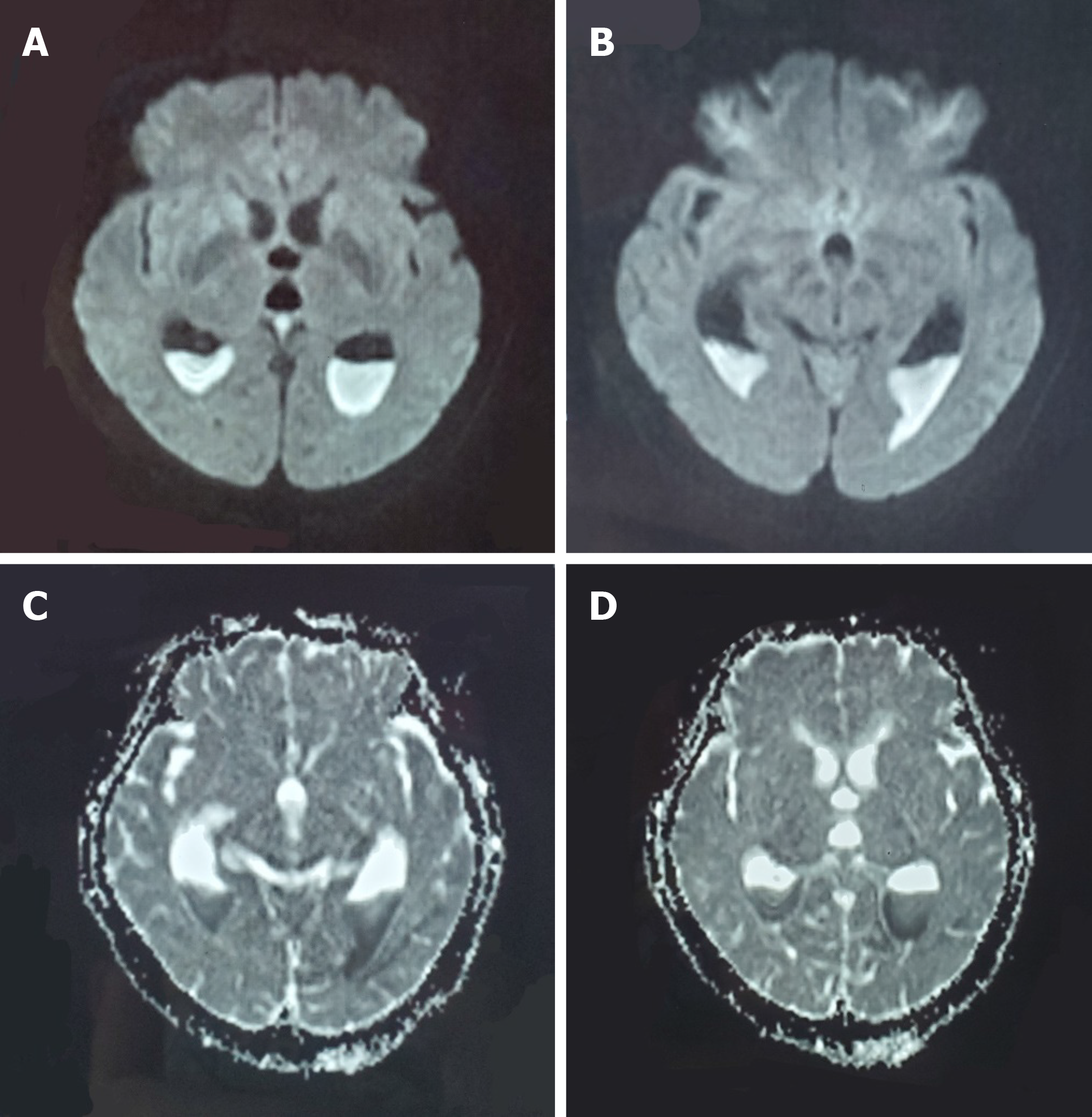
Figure 1 Head magnetic resonance imaging before admission.
A and B: The fester-traditional cerebrospinal fluid level was seen inside the posterior horns of the bilateral lateral ventricles, and the fester diffuse weighted imaging showed a high signal; C and D: The fester showed a low signal on the apparent diffusion coefficient map.
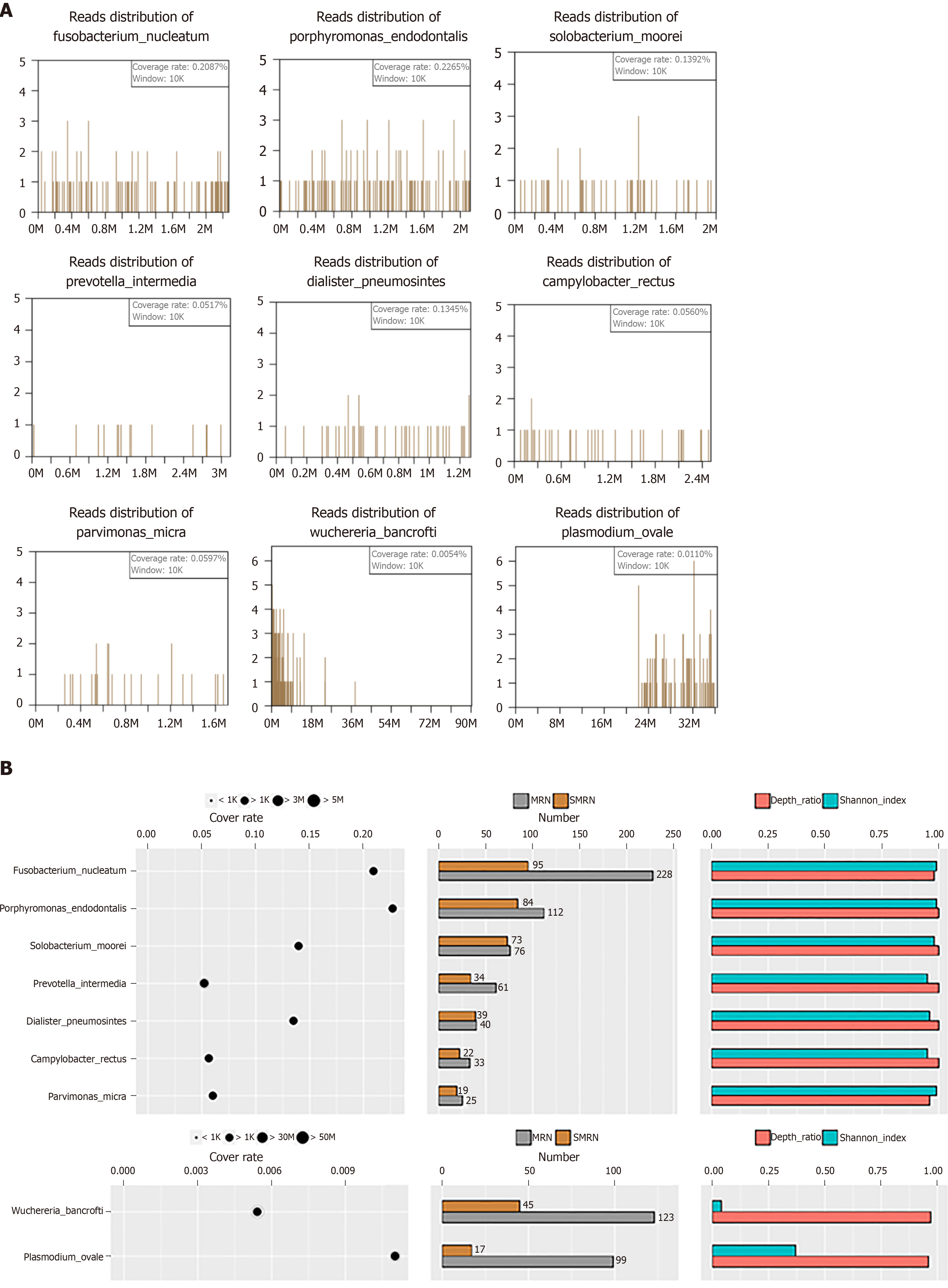
Figure 2 The next-generation sequencing results revealing the presence of the following pathogenic microorganisms in the traditional cerebrospinal fluid of the patient.
A and B: Fusobacterium, Porphyromonas, Solobacterium, Prevotella, Dialister, Fusobacterium nucleatum, Fusobacterium hwasookii, Porphyromonas endodontalis, Solobacterium moorei, Prevotella intermedia, and Dialister pneumosintes.
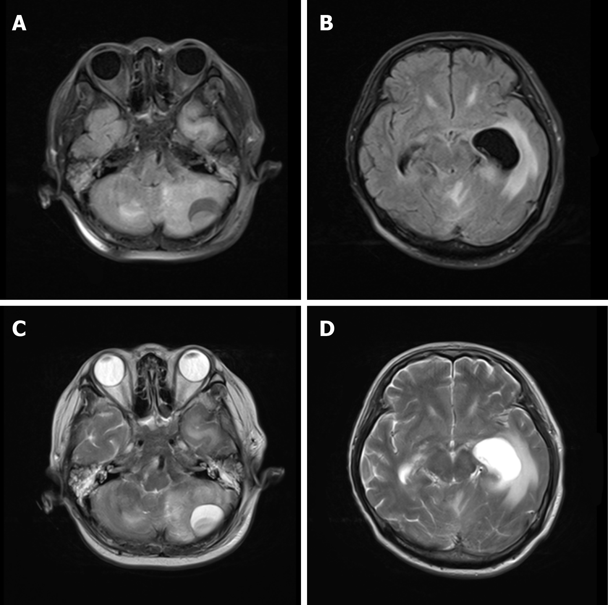
Figure 3 Head magnetic resonance imaging upon admission.
A-D: A quasicircular liquid signal opacity was seen in the left cerebellum, with a low T1WI signal (A) and a high T2WI signal (C). The adjacent brain stem was compressed, the fourth ventricle became narrower (A and B), and ventricular hydrops accompanied by paraventricular edema were observed.
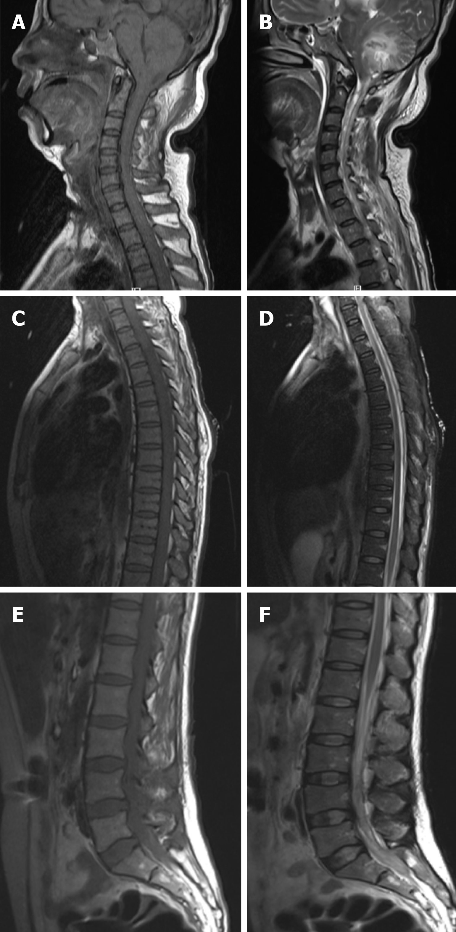
Figure 4 Spinal magnetic resonance imaging on admission.
A and B: Flake-like and stripe-like long T1 and long T2 signals were seen in the cerebellar hemisphere and cervical spinal cords on spinal magnetic resonance imaging (MRI); C and D: Long stripe T1 and T2 abnormal signals were seen in the T1-2 spinal cord on chest MRI; E and F: A flake-like low signal was seen in the spinal cord on lumbar MRI, and multiple nodular low signals were observed in the cauda equine innervation area at the L3-4 levels.
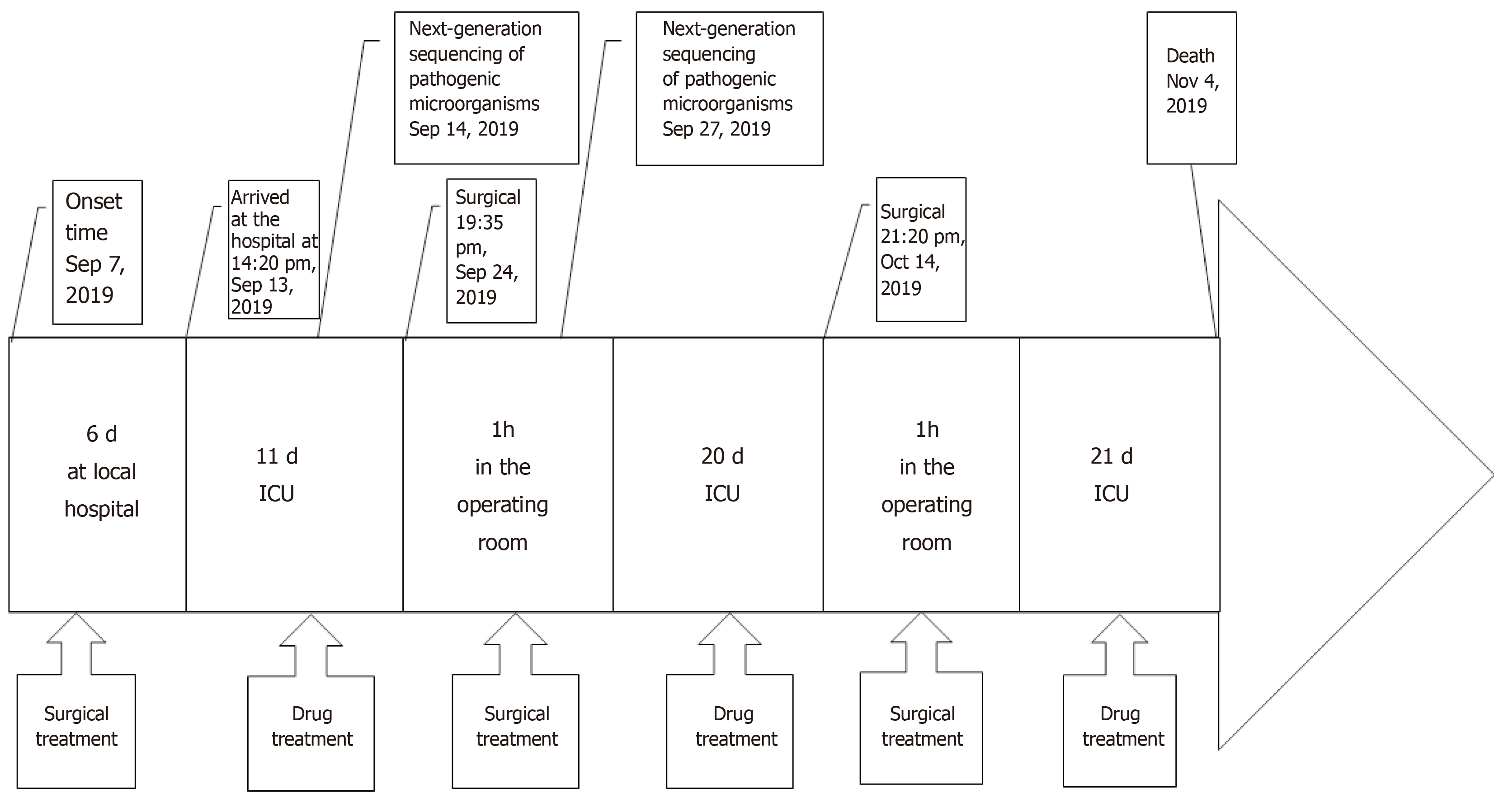
Figure 5 Timeline of the case.
ICU: Intensive care unit.
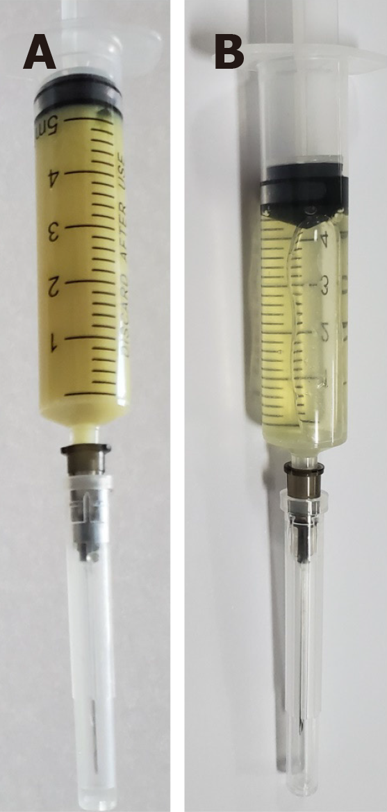
Figure 6 Traditional cerebrospinal fluid.
A: Traditional cerebrospinal fluid (CSF) on admission. The CSF was a rice water-like purulent liquid; B: CSF at 2 wk after admission. The CSF was a faint-yellow, clear liquid.
- Citation: Xue H, Wang XH, Shi L, Wei Q, Zhang YM, Yang HF. Dental focal infection-induced ventricular and spinal canal empyema: A case report. World J Clin Cases 2020; 8(14): 3114-3121
- URL: https://www.wjgnet.com/2307-8960/full/v8/i14/3114.htm
- DOI: https://dx.doi.org/10.12998/wjcc.v8.i14.3114


