Copyright
©The Author(s) 2020.
World J Clin Cases. Jul 26, 2020; 8(14): 3097-3107
Published online Jul 26, 2020. doi: 10.12998/wjcc.v8.i14.3097
Published online Jul 26, 2020. doi: 10.12998/wjcc.v8.i14.3097
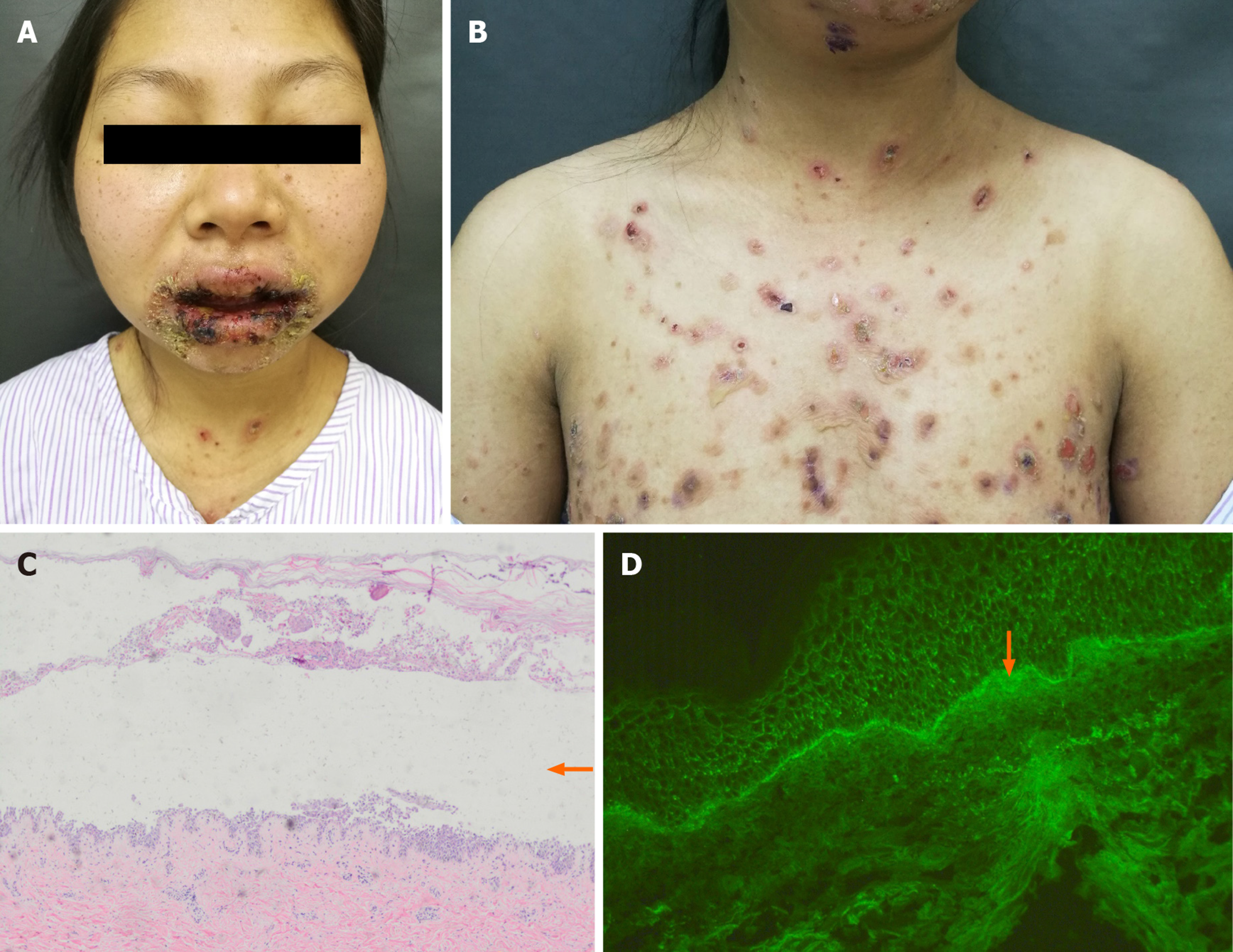
Figure 1 Diffuse lips and mucosal erythema and erosions (A), erythema and loose blisters on the trunk (B), intraepidermal acantholysis and blisters (orange arrow; HE × 40) (C), and linear deposition of C3 in the basal stratum of skin (orange arrow; × 100) (D).
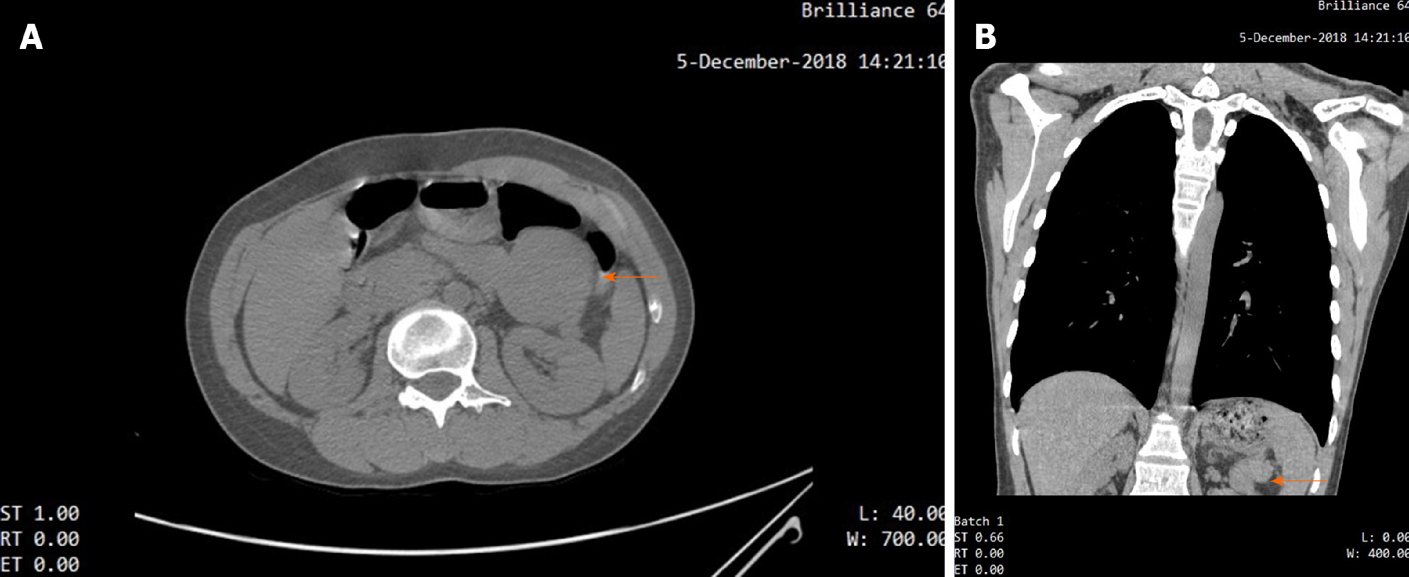
Figure 2 Computed tomography scan showed that there was an iso-dense, well-circumscribed mass (orange arrow) in the upper abdominal area.
A: Axial section; B: Coronal section.
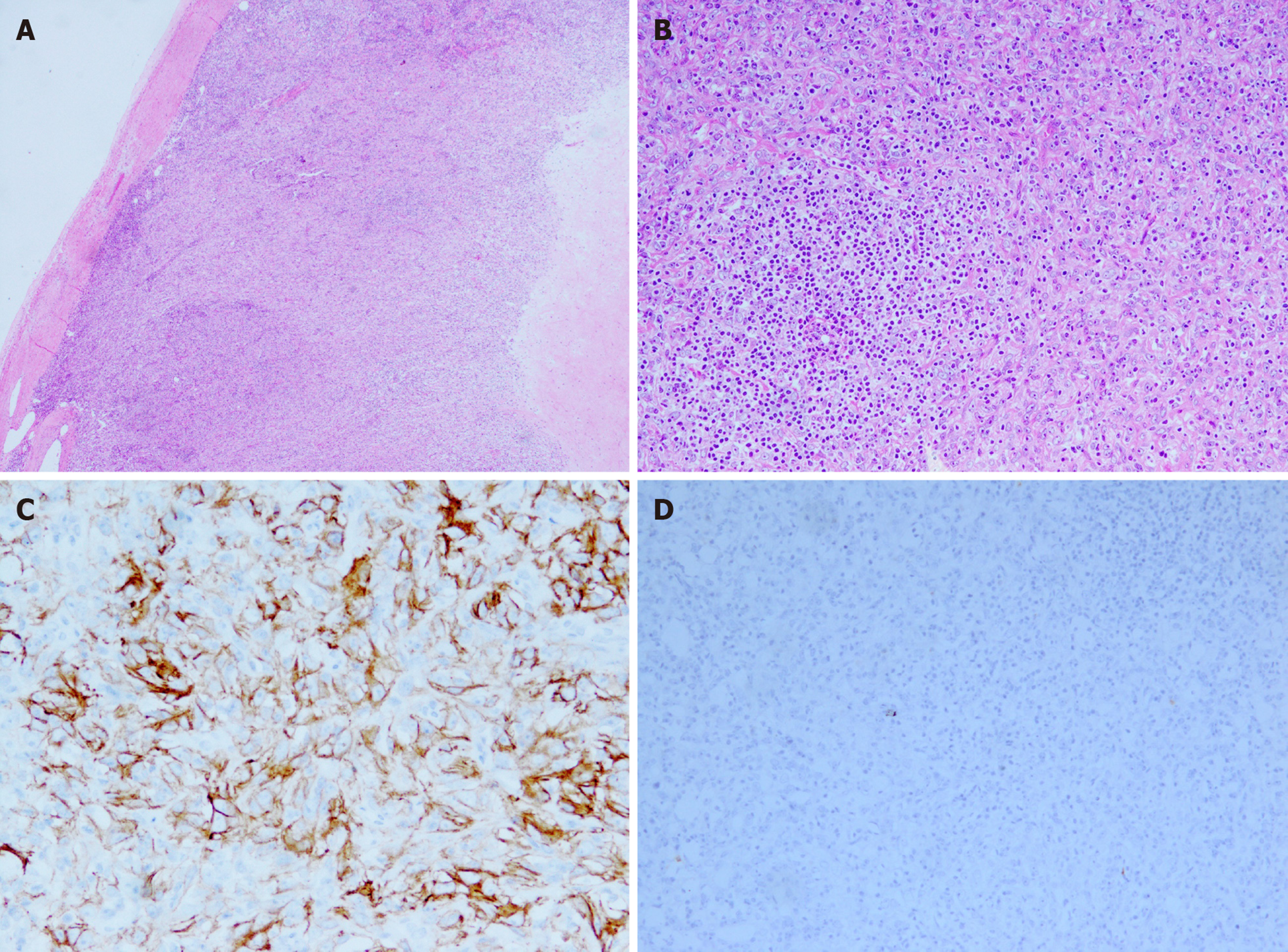
Figure 3 The tumor tissue had a clear boundary (HE × 20) (A), spindle tumour cells were distributed in the background of abundant small lymphocytes (HE × 100) (B), the neoplastic cells were strong positive for CD21 (× 200) (C), and the neoplastic cells were negative for CD117 (× 100) (D).
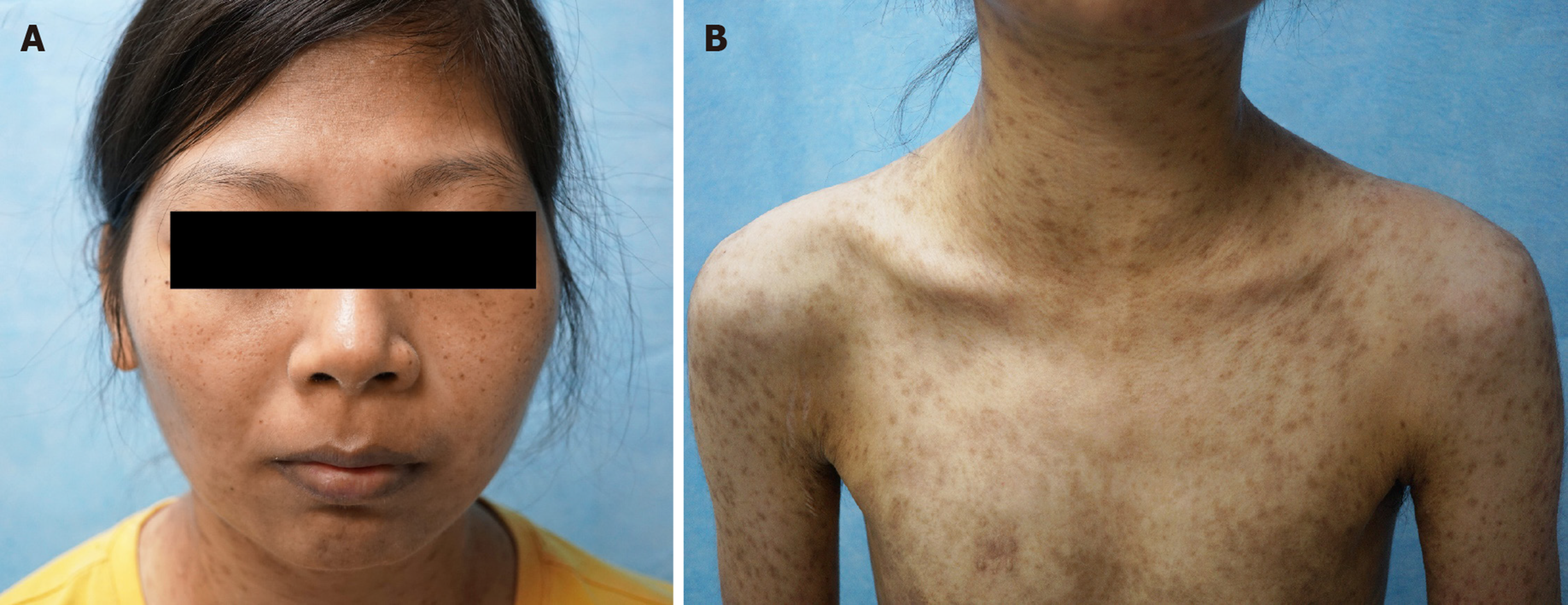
Figure 4 Mucosal and skin lesions had disappeared, leaving lichenoid hyperpigmentation behind (A and B).
- Citation: Zhuang JY, Zhang FF, Li QW, Chen YF. Intra-abdominal inflammatory pseudotumor-like follicular dendritic cell sarcoma associated with paraneoplastic pemphigus: A case report and review of the literature. World J Clin Cases 2020; 8(14): 3097-3107
- URL: https://www.wjgnet.com/2307-8960/full/v8/i14/3097.htm
- DOI: https://dx.doi.org/10.12998/wjcc.v8.i14.3097


