Copyright
©The Author(s) 2020.
World J Clin Cases. Jul 26, 2020; 8(14): 3090-3096
Published online Jul 26, 2020. doi: 10.12998/wjcc.v8.i14.3090
Published online Jul 26, 2020. doi: 10.12998/wjcc.v8.i14.3090
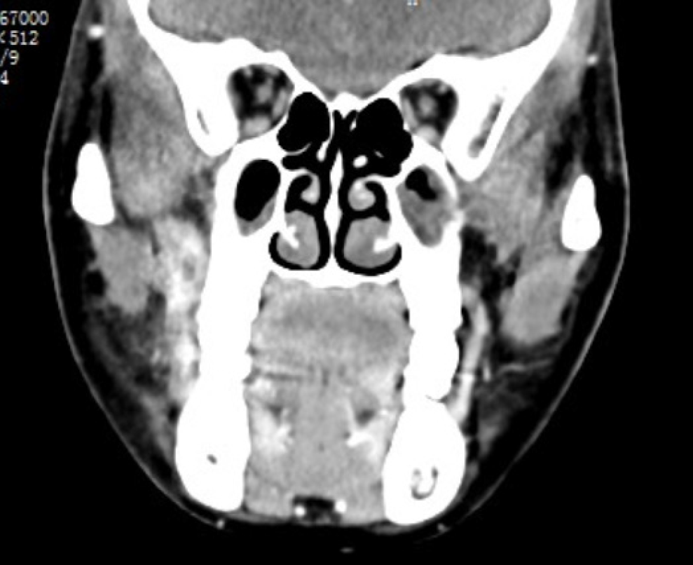
Figure 1 Computed tomography scan (axial section) showing an enhanced soft tissue mass in the right infratemporal fossa.
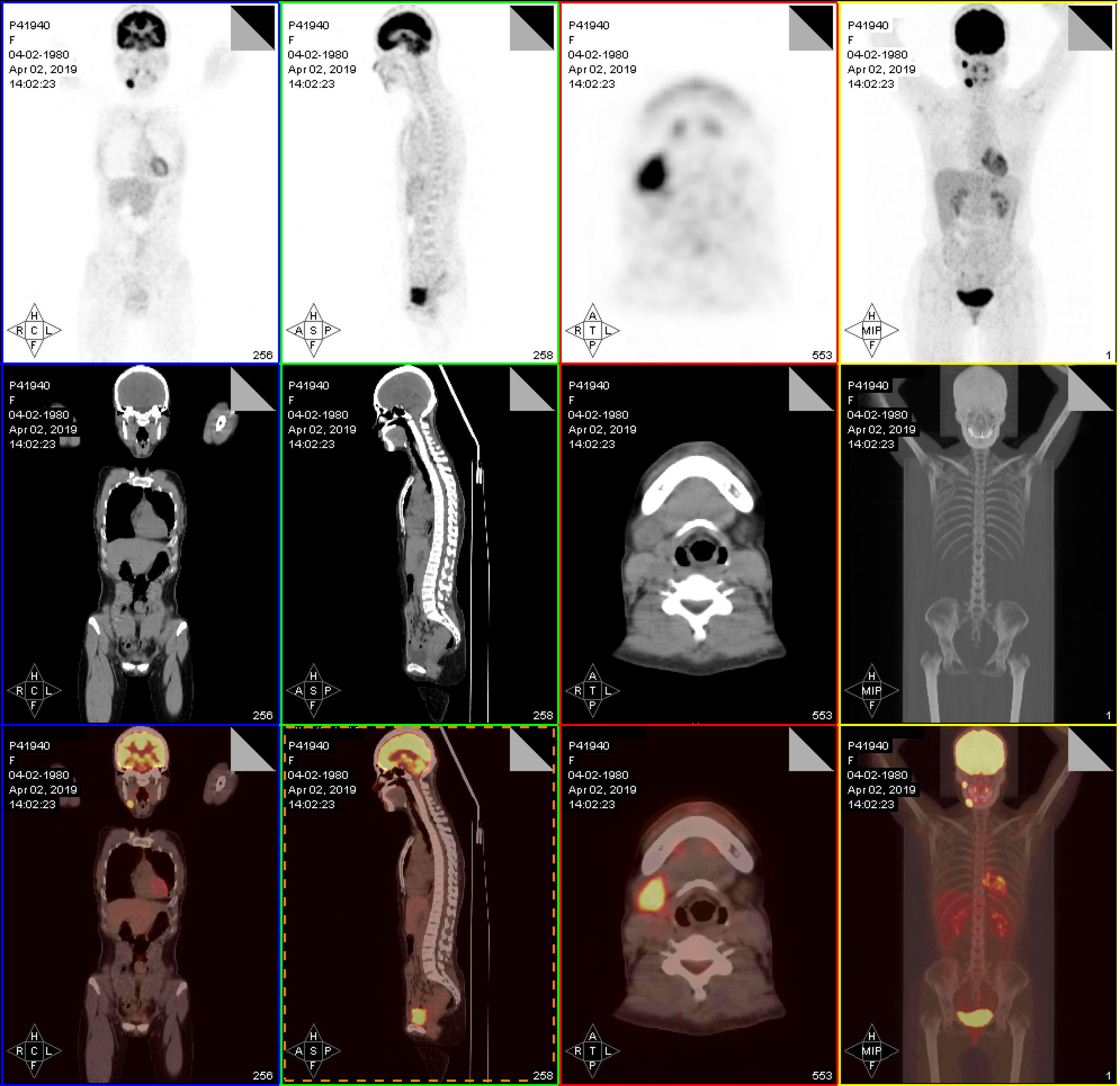
Figure 2 Representative fusion positron emission tomography/computed tomography images demonstrate intense uptake in the right infratemporal fossa and the lymph node of right II.
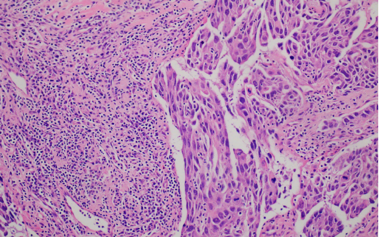
Figure 3 Hematoxylin-eosin staining of mucoepidermoid carcinoma.
High grade mucoepidermoid carcinoma: Infiltration and solid growth pattern (magnification: 200 ×).
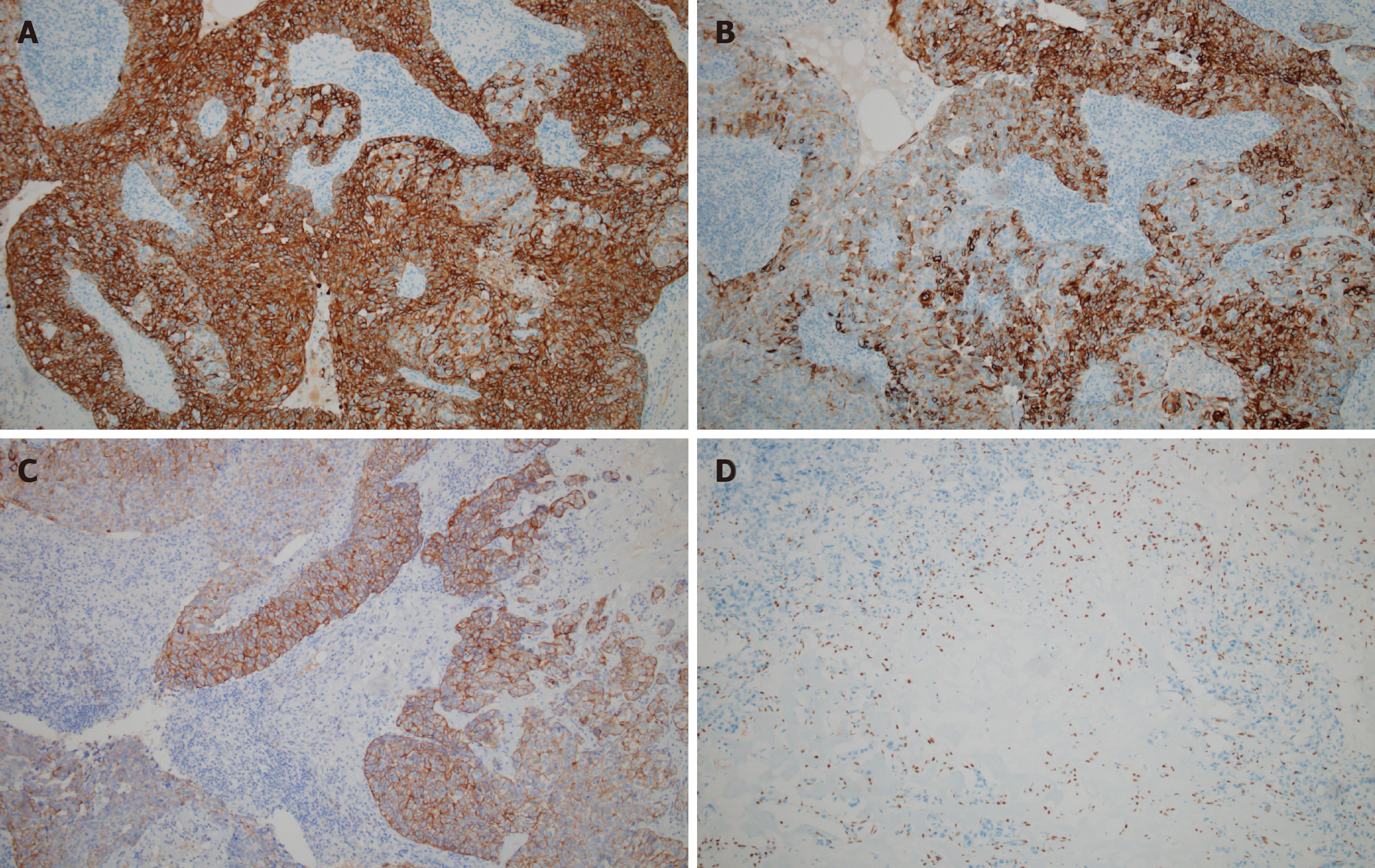
Figure 4 Immunophenotypic features of mucoepidermoid carcinoma.
A: Cytokeratin 7 showing diffusely positive staining; B: Showing positive staining for cytokeratin 5/6; C: Showing a strong positive reaction to cytokeratin 18; D: Focal positive expression of P40 (magnification: 200 ×).
- Citation: Zhang HY, Yang HY. Mucoepidermoid carcinoma in the infratemporal fossa: A case report. World J Clin Cases 2020; 8(14): 3090-3096
- URL: https://www.wjgnet.com/2307-8960/full/v8/i14/3090.htm
- DOI: https://dx.doi.org/10.12998/wjcc.v8.i14.3090


