Copyright
©The Author(s) 2020.
World J Clin Cases. Jul 26, 2020; 8(14): 3050-3056
Published online Jul 26, 2020. doi: 10.12998/wjcc.v8.i14.3050
Published online Jul 26, 2020. doi: 10.12998/wjcc.v8.i14.3050
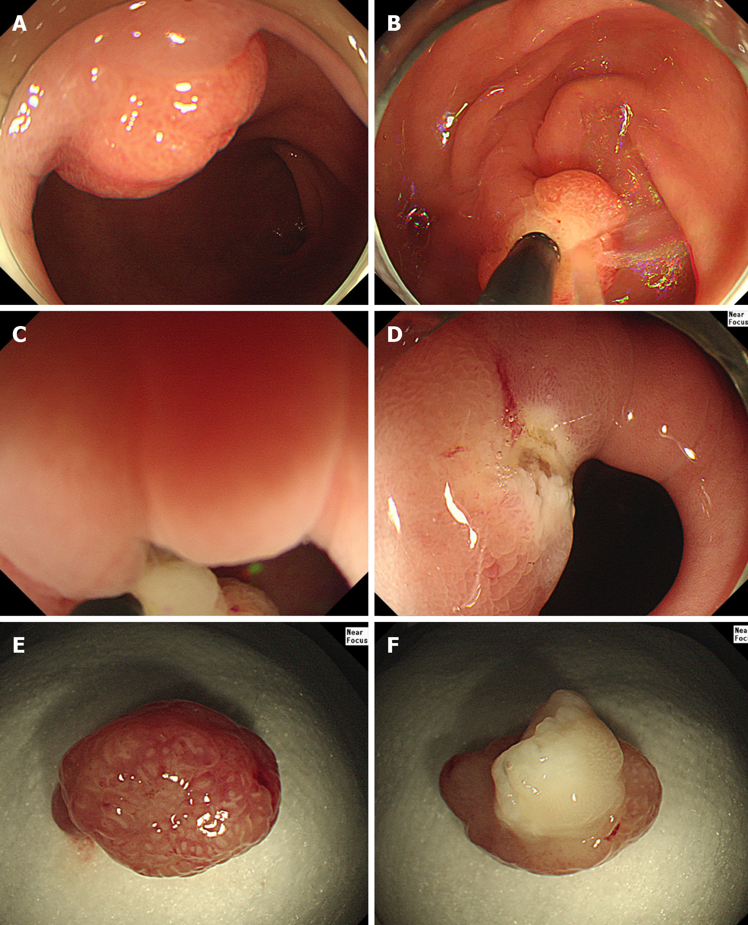
Figure 1 Underwater endoscopic mucosal resection in the first case.
A: Endoscopic view of the polyp in the pyloric ring; B: Filling water around the lesion; C: Snaring of the lesion in water; D: Endoscopic view of the resected area after endoscopic resection; E: The head portion of the resected polyp; F: The stalk portion of the resected polyp.
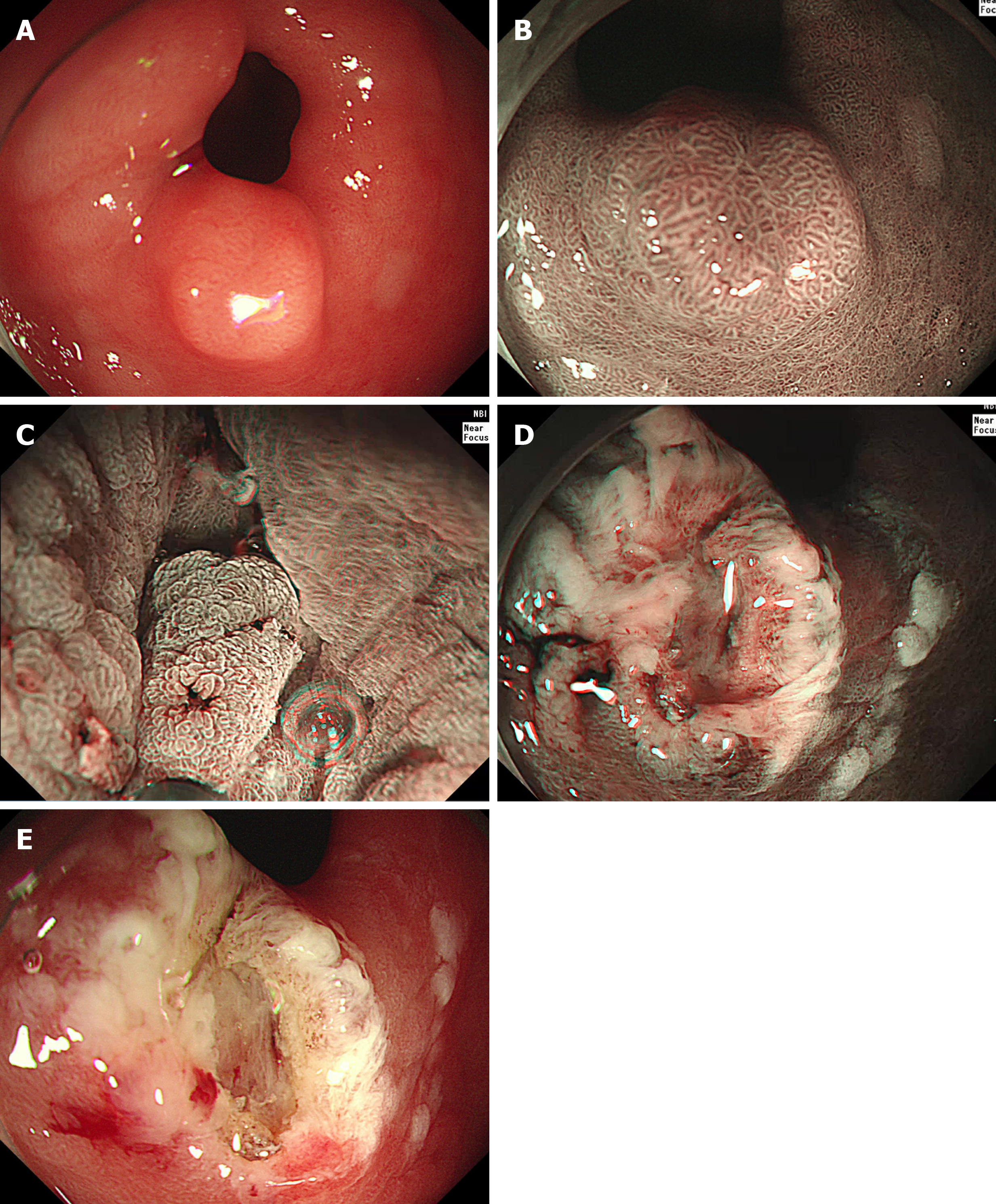
Figure 2 Underwater endoscopic mucosal resection in the third case.
A: Endoscopic view of the neoplasm in the pyloric ring; B: Endoscopic view of the neoplasm in the pyloric ring under narrow-band imaging; C: Snaring of the lesion in water under narrow-band imaging; D: Endoscopic view of the resected area after endoscopic resection under narrow-band imaging; E: Endoscopic view of the resected area after endoscopic resection.
- Citation: Kim DH, Park SY, Park CH, Kim HS, Choi SK. Underwater endoscopic mucosal resection for neoplasms in the pyloric ring of the stomach: Four case reports. World J Clin Cases 2020; 8(14): 3050-3056
- URL: https://www.wjgnet.com/2307-8960/full/v8/i14/3050.htm
- DOI: https://dx.doi.org/10.12998/wjcc.v8.i14.3050


