Copyright
©The Author(s) 2020.
World J Clin Cases. Jul 26, 2020; 8(14): 3039-3049
Published online Jul 26, 2020. doi: 10.12998/wjcc.v8.i14.3039
Published online Jul 26, 2020. doi: 10.12998/wjcc.v8.i14.3039
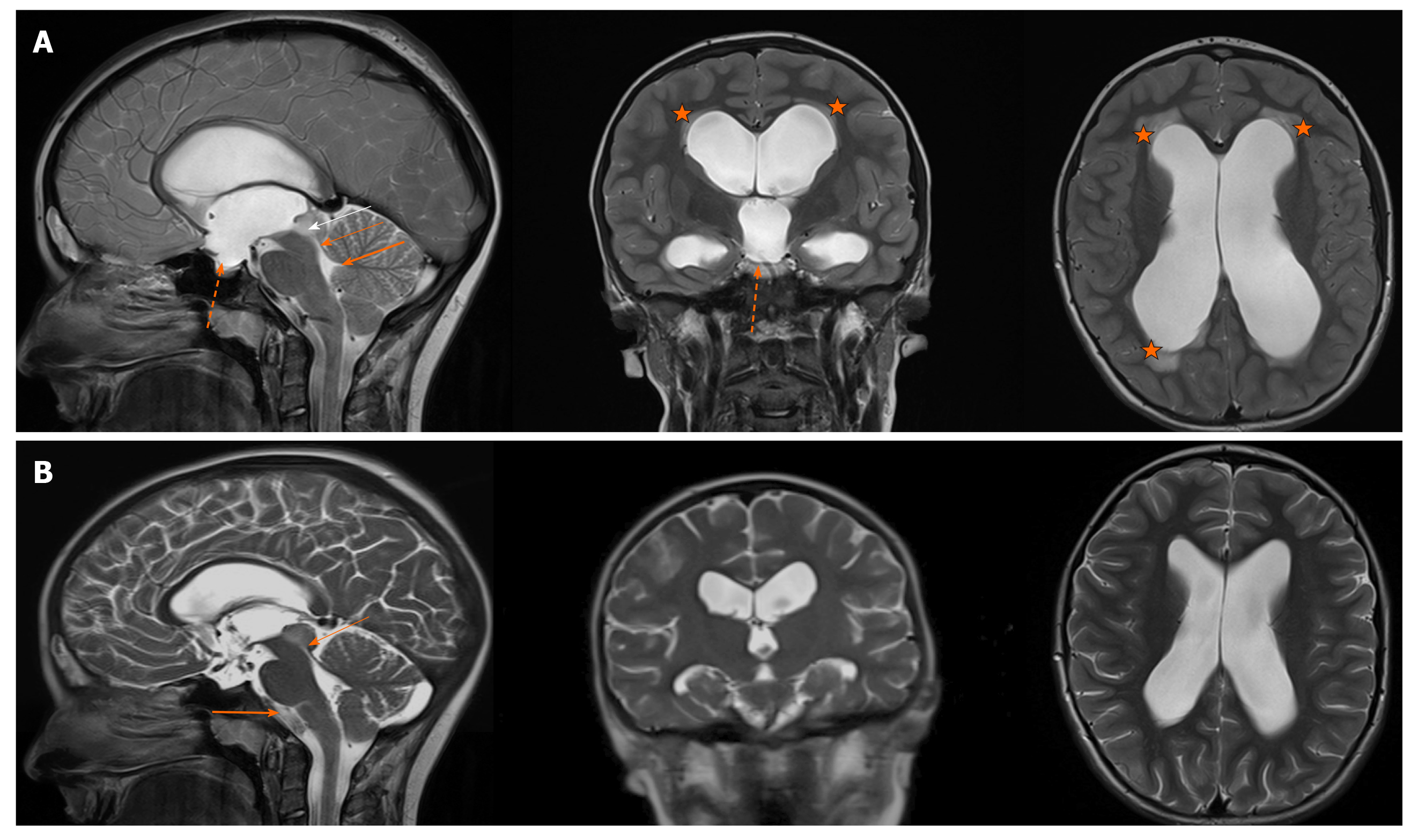
Figure 1 The T2-phases of the magnetic resonance imaging (sagittal, coronal and axial views) revealing a tumour in the right part of the tectum (white arrow), obstructing the aqueduct of Sylvius and causing extensive hydrocephalus.
A: The enlarged third ventricle is bulging into the sella turcica (dotted arrow). Periventricular lucences can also be seen (star). The thick arrow indicates the stenosis, the thin arrow indicates a normal fourth ventricle width. B: After one year, the control MRI (T2-phase) showed the regression of ventriculomegaly as a result of the endoscopic third ventriculostomy (ETV). The stenosis in the aqueduct is still present to the same degree (thin arrow). The cerebrospinal fluid flow artefact in the third ventricle can be seen (thick arrow), confirming the third ventricle floor opening after the successful ETV. The tumour has not been progressing. The supratentorial ventricles are narrower.
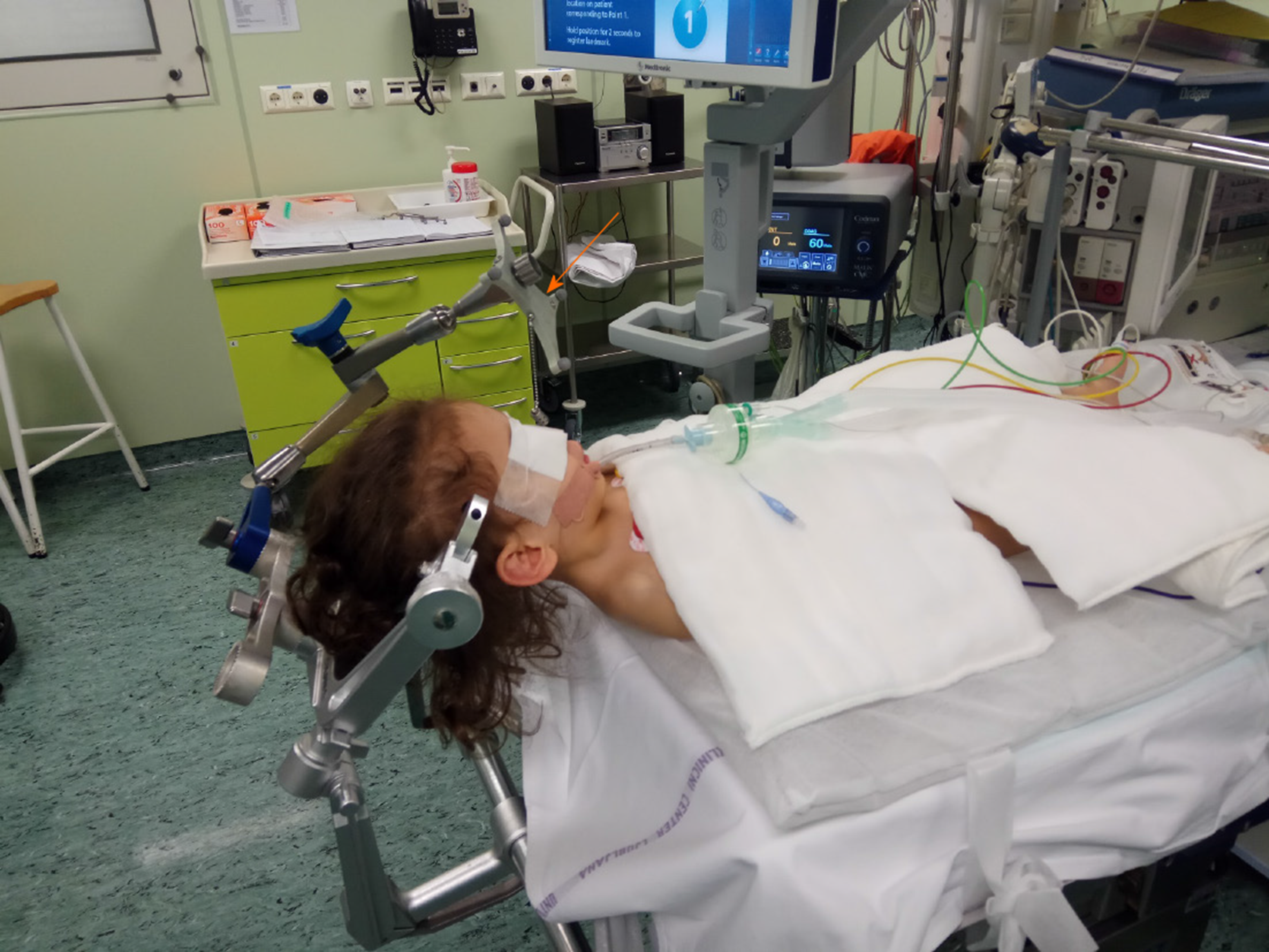
Figure 2 The patient in a supine position under general anaesthesia, prepared for the endoscopical approach.
The head is elevated 20 degrees and the neck is slightly flexed. The neuronavigation reference point (antenna) for guided neuroendoscopy can be seen on the Mayfield frame (arrow).
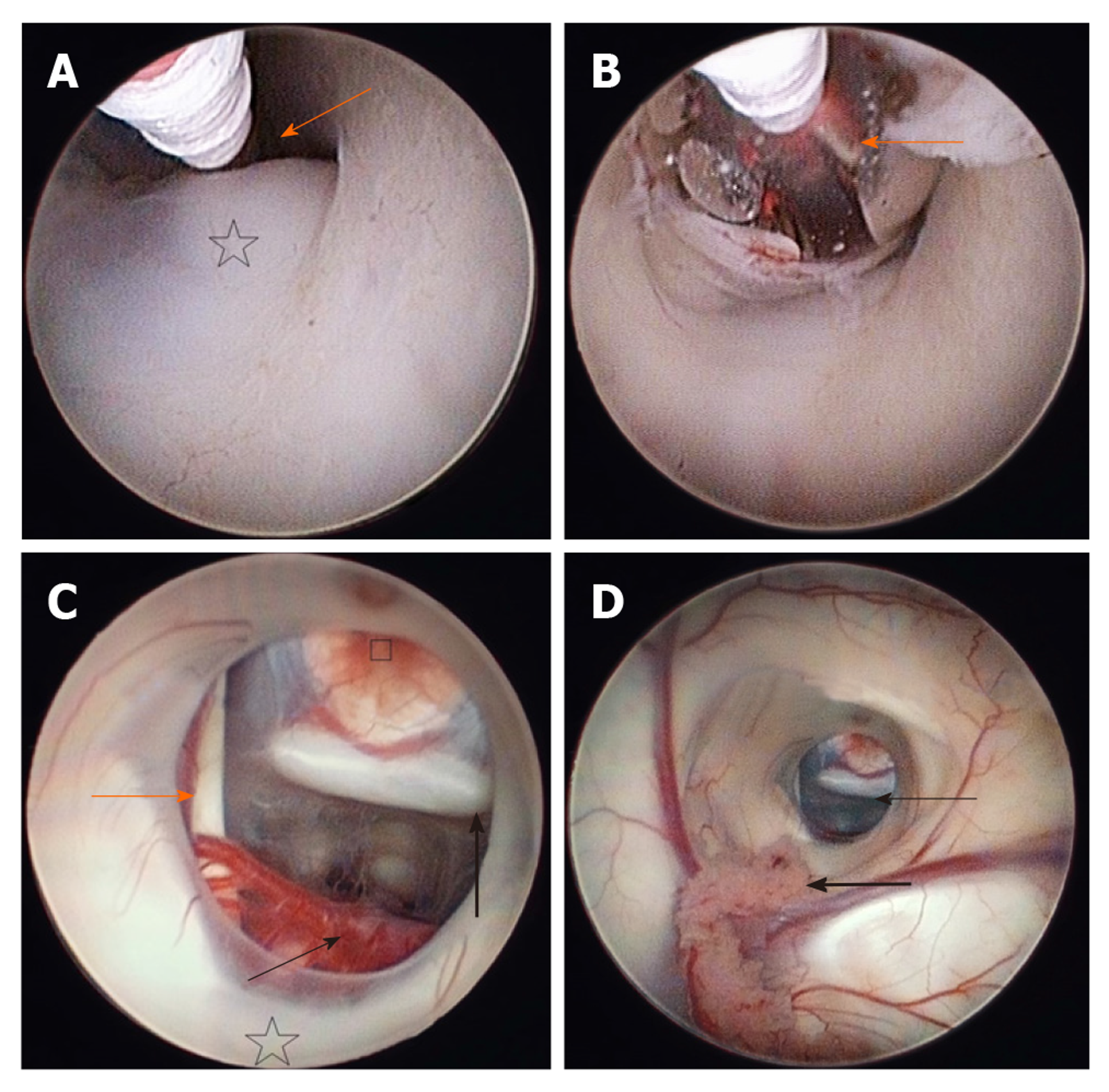
Figure 3 The floor of the third ventricle.
A: The tip of the Fogarty balloon is seen, located in the area of the thin, translucent membrane, just in front of the mammillary bodies (star), which is the target for the fenestration (arrow); B: The fenestration is underway. The floor of the third ventricle has just ben perforated and the opening is being enlarged with a Fogarty balloon. The vessels of the posterior circulation can be seen through the translucent wall of the inflated balloon (arrow); C: The ventriculostomy after the completed fenestration. The posterior circulation (the basilar artery with its bifurcation into posterior cerebral arteries and their branches) can be seen posteriorly (thin arrow), anteriorly is the clivus, the dorsum sellae above (thick arrow) and the tuber cinereum (square). The 3rd cranial nerve is running on the left (orange arrow). The furthest behind, just above the fenestration, mammillary bodies and thalami are located (star); D: The fenestration seen from the lateral ventricle through the foramen of Monro. In the opening, the clivus can be clearly seen (thin arrow) and the tuber cinereum in the front. The choroid plexus is located at the bottom of the figure (thick arrow).
- Citation: Munda M, Spazzapan P, Bosnjak R, Velnar T. Endoscopic third ventriculostomy in obstructive hydrocephalus: A case report and analysis of operative technique. World J Clin Cases 2020; 8(14): 3039-3049
- URL: https://www.wjgnet.com/2307-8960/full/v8/i14/3039.htm
- DOI: https://dx.doi.org/10.12998/wjcc.v8.i14.3039


