Copyright
©The Author(s) 2020.
World J Clin Cases. Jul 26, 2020; 8(14): 2902-2916
Published online Jul 26, 2020. doi: 10.12998/wjcc.v8.i14.2902
Published online Jul 26, 2020. doi: 10.12998/wjcc.v8.i14.2902
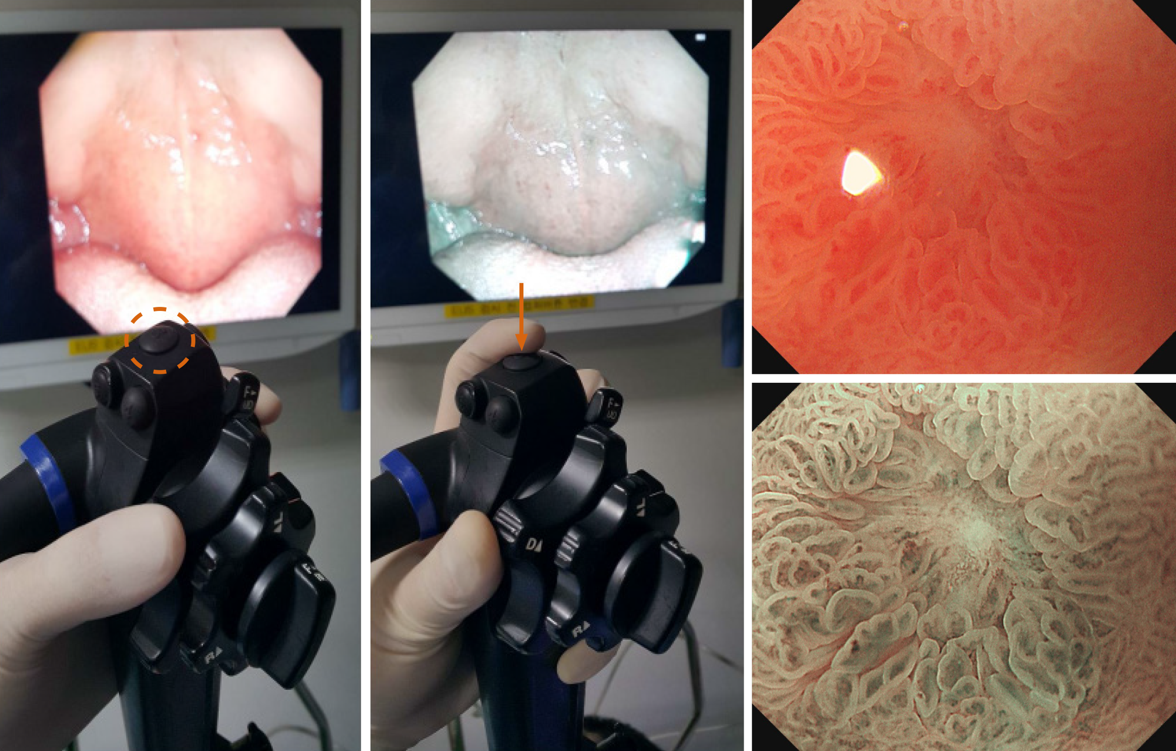
Figure 1 Narrow-band imaging system.
When the button is pressed by the endoscopist, the endoscopy monitor changes from white-light imaging to narrow-band imaging (NBI) mode. Compared to the magnification mode without NBI (upper-right panel), NBI enables examination of the mucosal surface and microvessels around the erosion (lower-right panel).
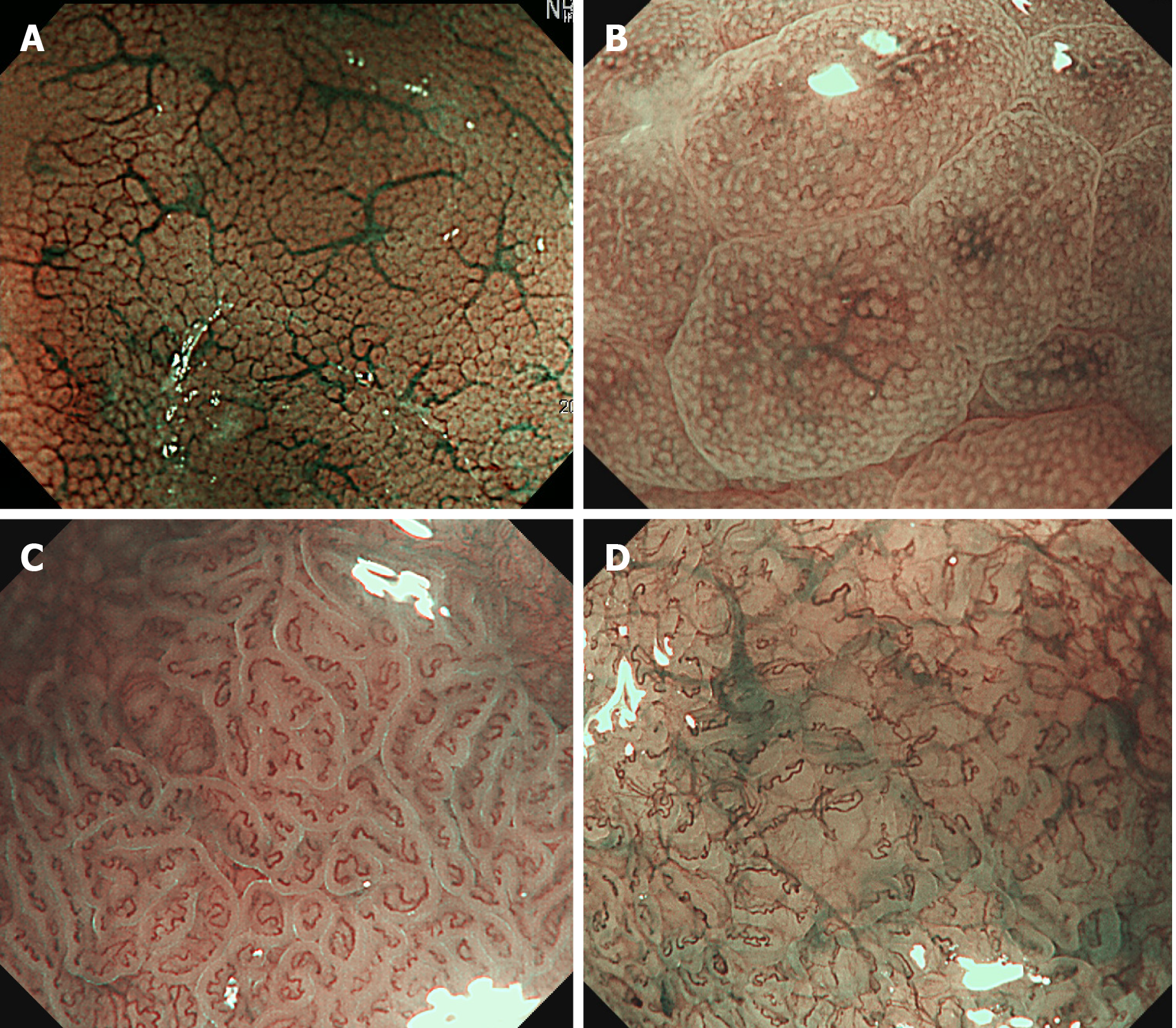
Figure 2 Magnifying narrow-band imaging endoscopy images of Helicobacter pylori-positive corpus mucosa and atrophic gastritis.
A: Normal mucosal pattern showing a honeycomb-like subepithelial capillary network and regular arrangement of collecting venules; B: Helicobacter pylori-infected status presenting as polygonal swollen mucosa with dilated round crypt opening; C: Ridged surface structures encasing dilated coiled subepithelial capillaries indicate the presence of atrophic gastritis; D: Severe mucosal atrophy characterized by irregular coiled microvessels, loss of gastric pits, and greenish submucosal vessels.
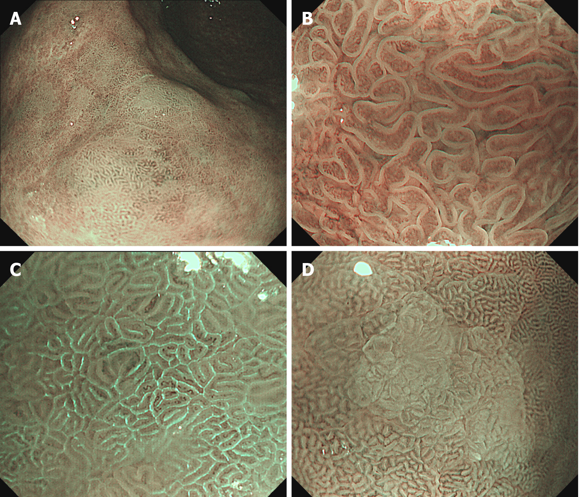
Figure 3 Narrow-band imaging endoscopy images of intestinal metaplastic mucosa.
A: Gastric mucosa covered with multiple whitish green-colored patches; B: Thick borders enclosing a tubulovillous mucosal pattern, the so-called marginal turbid band (MTB); C: Thin fluorescent lines along the MTB, the so-called light blue crest; D: White opaque substance obscuring the mucosa surface.
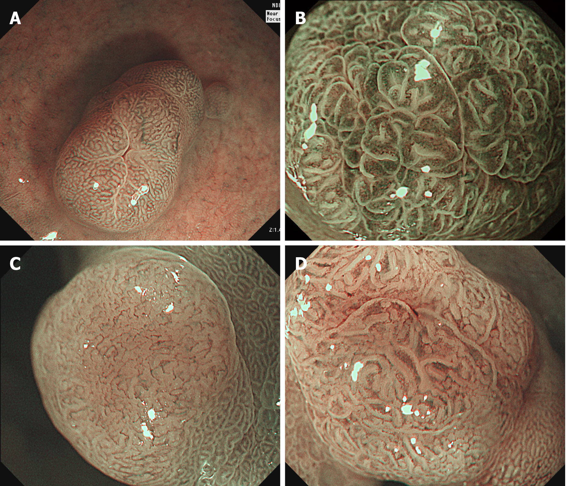
Figure 4 Magnifying narrow-band imaging findings of microvascular patterns for diagnosing the gastric polypoid lesions.
A: Honeycomb-like pattern (fundic gland polyp); B: Dense vascular pattern (hyperplastic polyp); C: Fine network within a light brown area (low grade dysplasia); D: Core vascular pattern (low grade dysplasia).
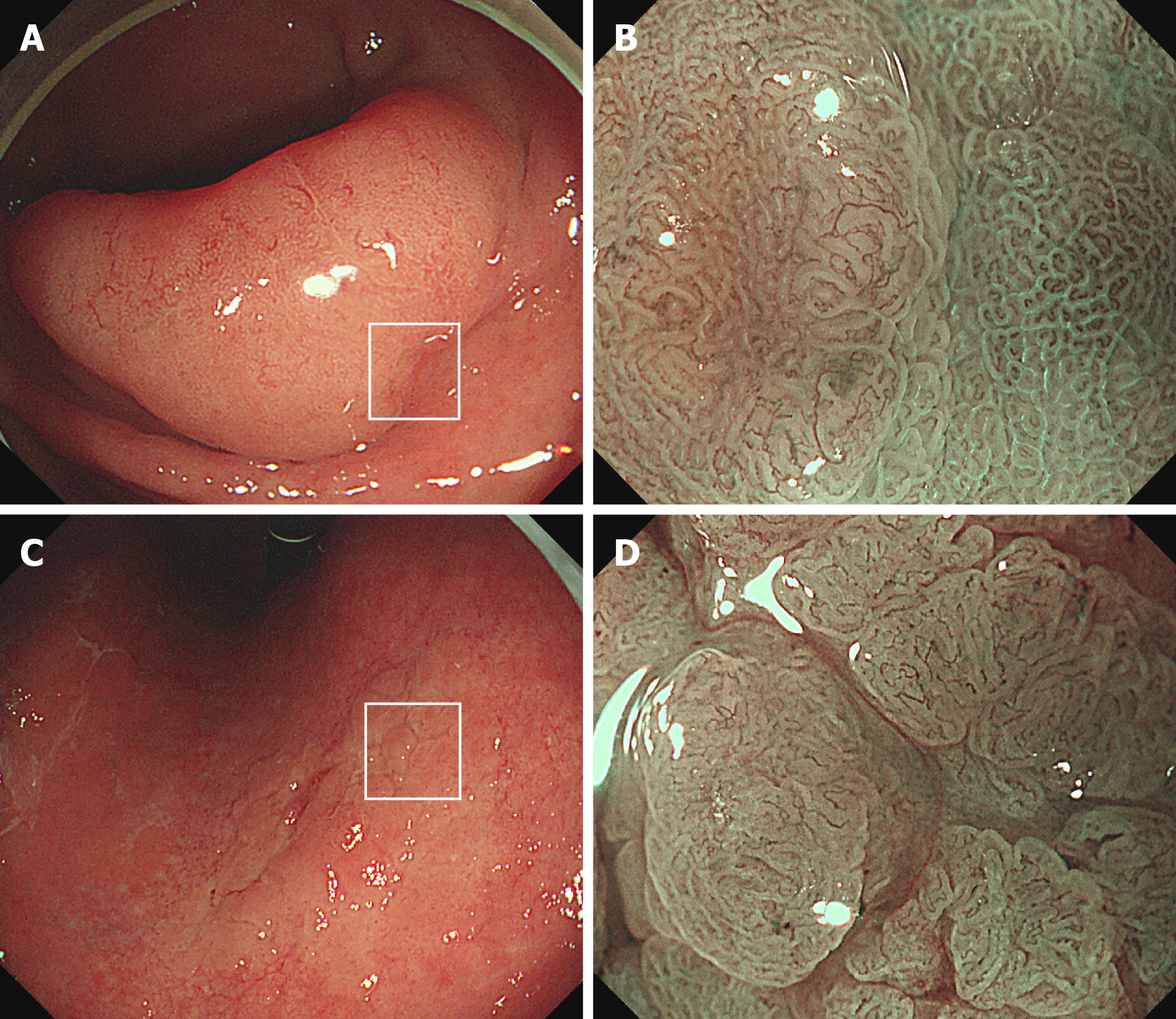
Figure 5 White-light endoscopy and magnifying narrow-band imaging images of high grade dysplasia.
A: An elevated lesion (40 mm × 30 mm) at the gastric antrum; B: Brownish area showing an irregular mucosal surface and a microvascular pattern (left side), indicating a high grade dysplasia. The demarcation line is evident between the dysplasia and background mucosa with intestinal metaplasia (also see white box in A); C: A slightly elevated lesion (35 mm × 20 mm) at the lesser curvature of the gastric corpus; D: The microvessels within irregular, nodular lesion show tortuosity and variation in shape. This magnifying narrow-band imaging endoscopic finding indicates high grade dysplasia (also see white box in C).
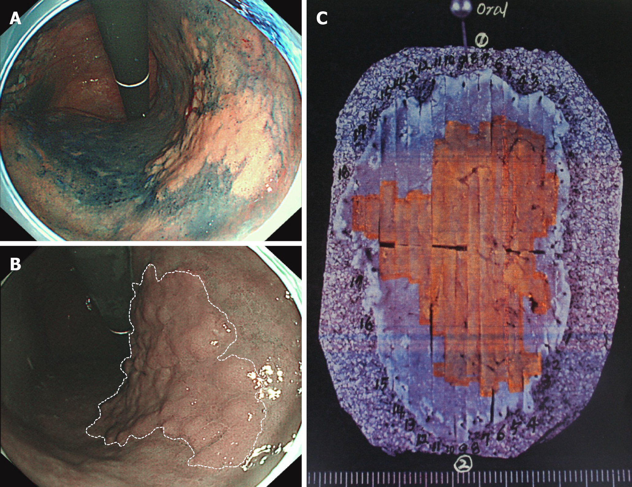
Figure 6 Narrow-band imaging endoscopy for determining the horizontal margin of gastric dysplasia before endoscopic submucosal dissection.
A: Conventional chromoendoscopy using indigo carmine is useful for determining the horizontal margin of gastric neoplasia. However, this procedure requires a dye solution and is time-consuming; B: When the endoscopist presses the button, narrow-band imaging (NBI) endoscopy can be easily performed as a virtual chromoendoscopy. The tumor margin is evident between the large brownish lesion and greenish background mucosa (dotted white line); C: An en bloc-resected specimen by endoscopic submucosal dissection. The orange-colored area indicates a tubulovillous adenoma of 46 mm × 36 mm size, corresponding to the tumor extent determined by NBI endoscopy.
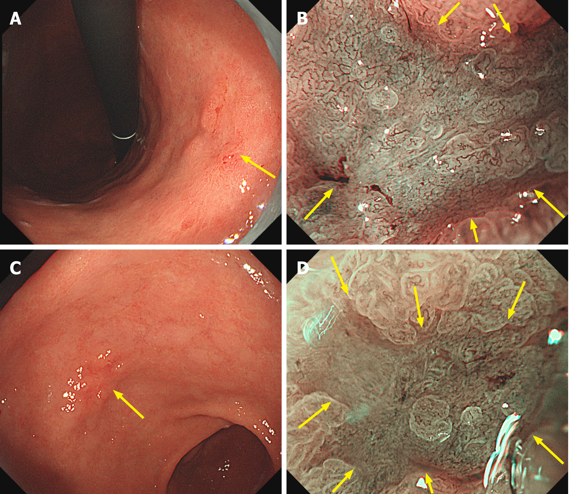
Figure 7 Magnifying narrow-band imaging endoscopic findings of early gastric cancers.
A: Conventional white-light endoscopy shows a slightly elevated and depressed lesion at the gastric corpus; B: Magnifying narrow-band imaging (NBI) endoscopy demonstrates an irregular microvascular and microsurface pattern with a demarcation line (yellow arrows); C: Conventional white-light endoscopy shows a reddish depressed lesion at the gastric antrum; D: By magnifying NBI endoscopy, an irregular microsurface pattern is identified within the demarcation line (yellow arrows).
- Citation: Cho JH, Jeon SR, Jin SY. Clinical applicability of gastroscopy with narrow-band imaging for the diagnosis of Helicobacter pylori gastritis, precancerous gastric lesion, and neoplasia. World J Clin Cases 2020; 8(14): 2902-2916
- URL: https://www.wjgnet.com/2307-8960/full/v8/i14/2902.htm
- DOI: https://dx.doi.org/10.12998/wjcc.v8.i14.2902


