Copyright
©The Author(s) 2020.
World J Clin Cases. Jul 6, 2020; 8(13): 2862-2869
Published online Jul 6, 2020. doi: 10.12998/wjcc.v8.i13.2862
Published online Jul 6, 2020. doi: 10.12998/wjcc.v8.i13.2862
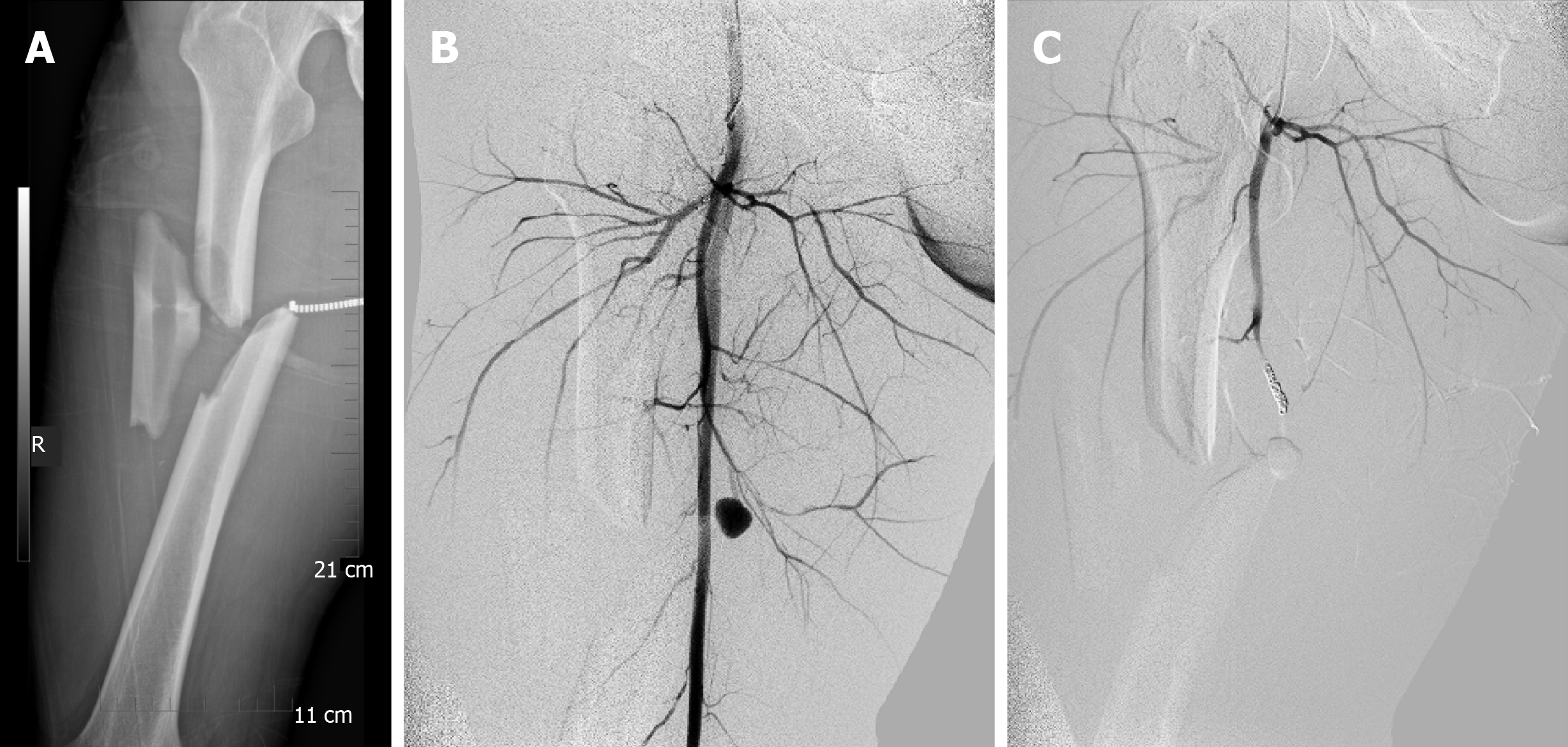
Figure 1 Imaging examinations.
A: Anteroposterior images of right femoral when the patient was delivered to the hospital; B: Computed tomography angiography showed accumulation of the contrast agents at the branch of femoral deep artery, indicating the rupture of the artery; C: A coil was used to embolize the ruptured artery.
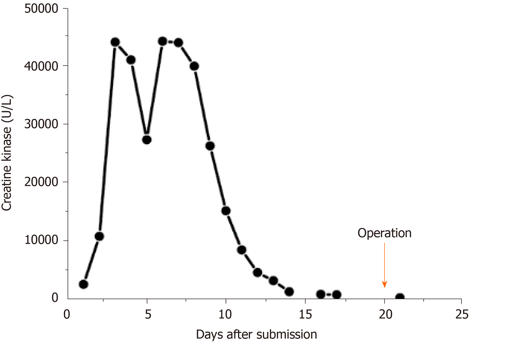
Figure 2 Creatine kinase change in the patient.
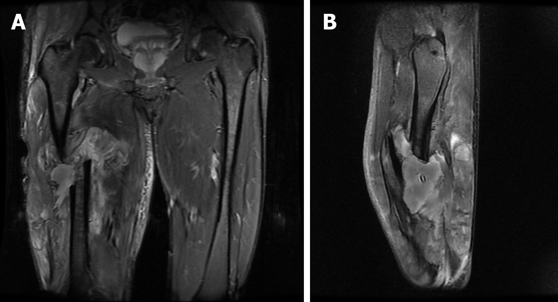
Figure 3 Preoperative magnetic resonance imaging (short TI inversion recovery) of lower limbs showed that the middle part of the right femoral shaft was discontinuous, the stump was dislocated, and the surrounding muscles were swollen.
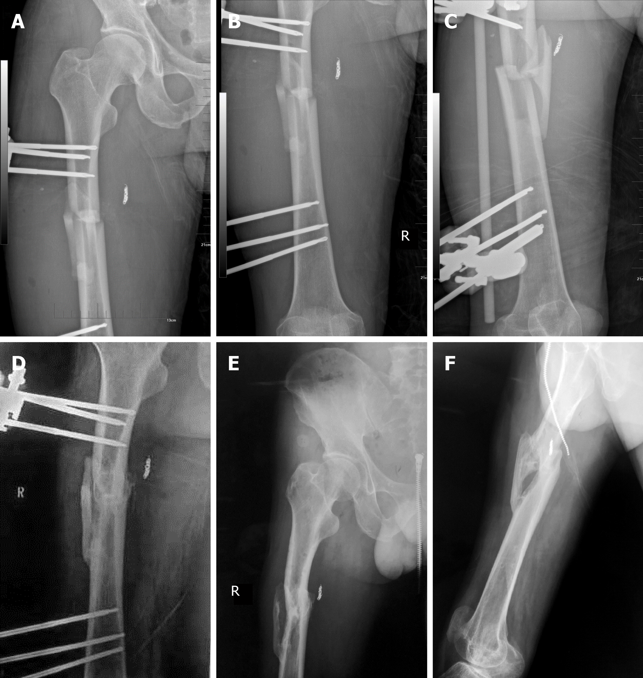
Figure 4 External fixation for 1 yr and 3 yr of follow-up showed improvement of the patient’s overall conditions and muscle strength.
A-C: Images of right femoral 1 d postoperatively; D: 1-yr follow-up at the local hospital showed that the fracture line had been blurred with mass formation of callus at the fracture site; E and F: 3-yr follow-up radiography revealed union of the fracture.
- Citation: Ge J, Kong KY, Cheng XQ, Li P, Hu XX, Yang HL, Shen MJ. Missed diagnosis of femoral deep artery rupture after femoral shaft fracture: A case report. World J Clin Cases 2020; 8(13): 2862-2869
- URL: https://www.wjgnet.com/2307-8960/full/v8/i13/2862.htm
- DOI: https://dx.doi.org/10.12998/wjcc.v8.i13.2862


