Copyright
©The Author(s) 2020.
World J Clin Cases. Jun 26, 2020; 8(12): 2647-2654
Published online Jun 26, 2020. doi: 10.12998/wjcc.v8.i12.2647
Published online Jun 26, 2020. doi: 10.12998/wjcc.v8.i12.2647
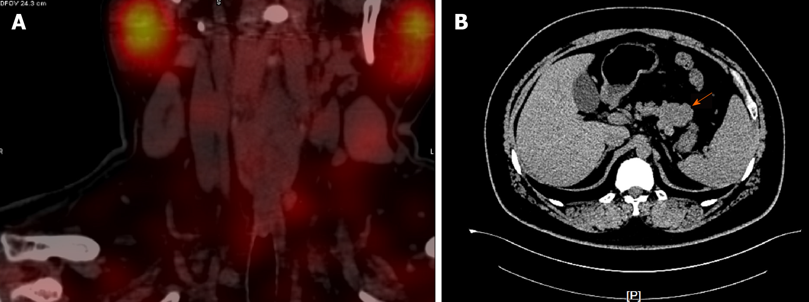
Figure 1 Computed tomography examination.
A: Parathyroid two-phase computed tomography revealed a strong signal in the upper part of the left lobes and posterior part of the right lobes of the thyroid; B: Pancreatic perfusion computed tomography imaging revealed irregular soft tissue densities of the pancreas body, which were localized around the stomach wall.
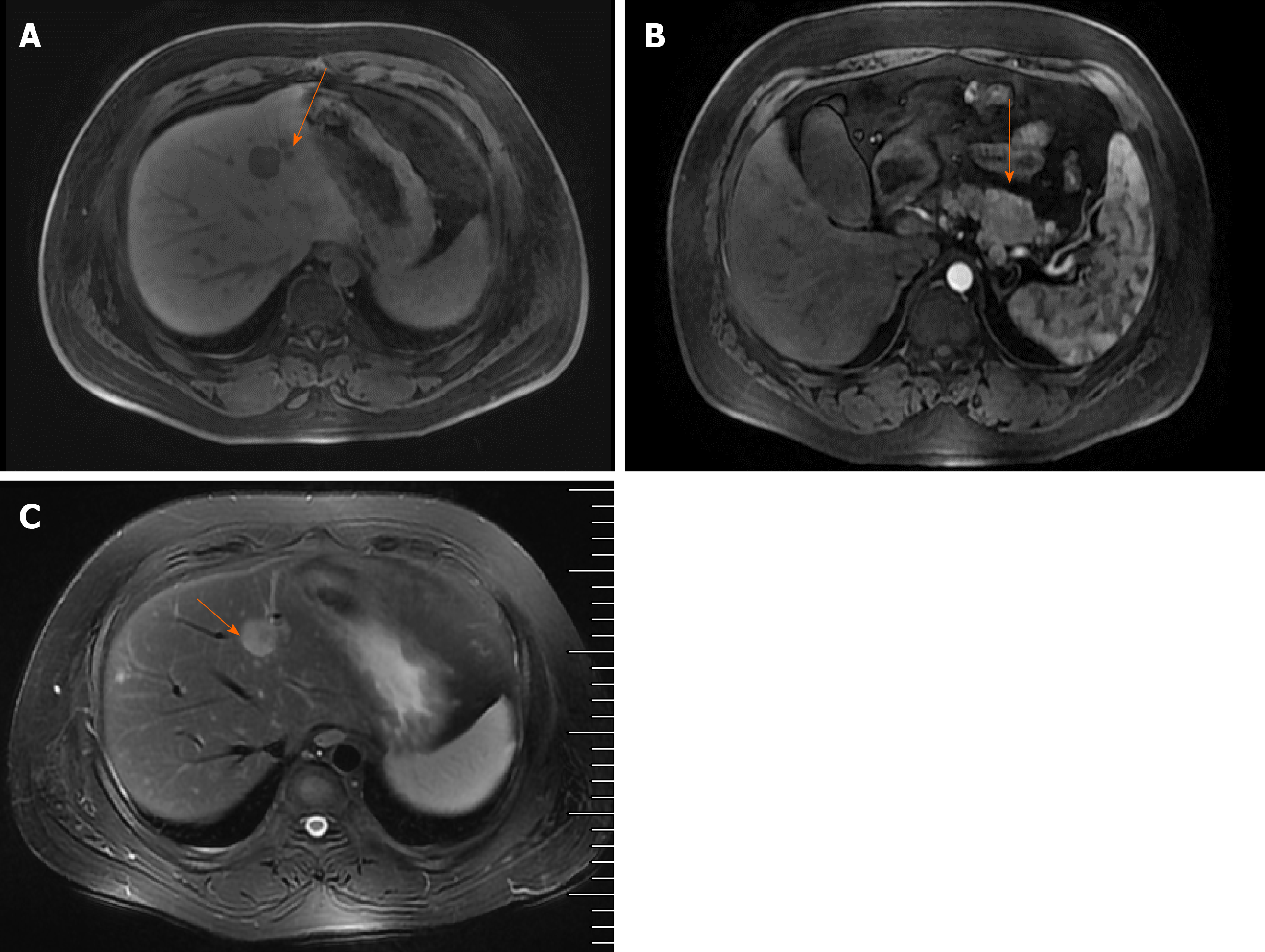
Figure 2 resonance imaging (MRI) examination revealed multiple abnormal signals in the liver segment II, segment VIII, and the junction area of liver segment II and IV as well as occupied lesions in the tail of the pancreatic body; B: Enhanced MRI revealed occupied lesions in the body and tail of the pancreas; C: Enhanced MRI revealed that the liver segment IV was occupied, suggesting the possibility of angiomyolipoma.
There were also small cysts in segments II and VIII of the liver and occupied lesions in the tail of the pancreatic body.
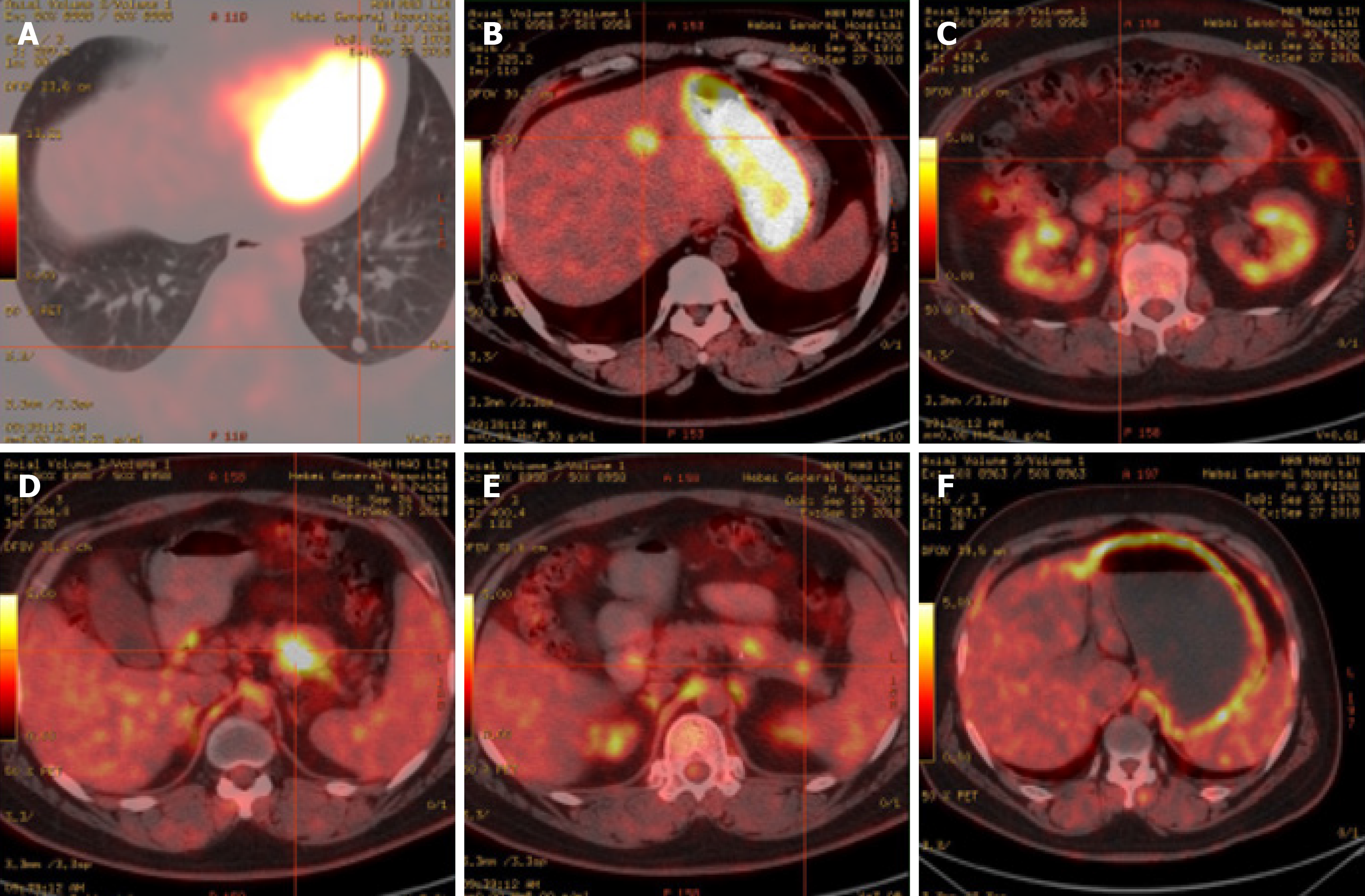
Figure 3 Positron emission tomography/computed tomography examination.
A, B, D and E: Hypermetabolic lesions in the lungs (A), intrahepatic segment IV (B), and the body (D) and tail (E) of the pancreas; C: No metabolic round nodules in the anterior pancreas; F: Diffusely increased metabolism in the stomach wall.
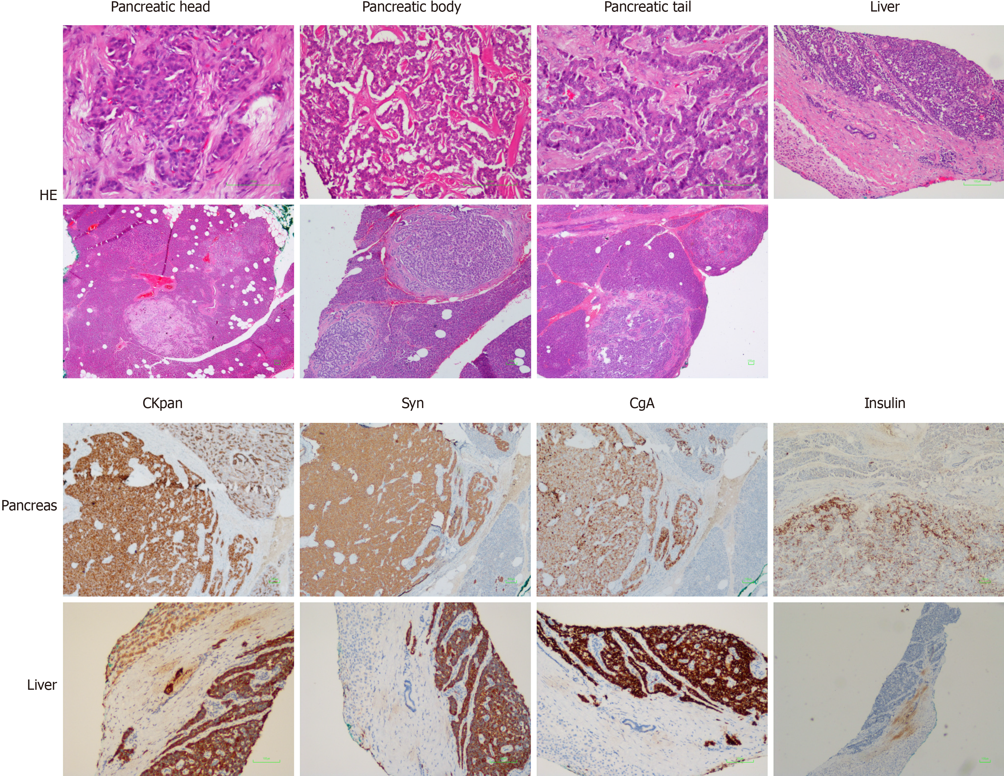
Figure 4 Pathological examination.
Hematoxylin & eosin staining showed (magnification, 40 ×; 100 ×) multiple nodules next to the pancreatic tumor, in the pancreatic tissue around the pancreatic body, and next to the pancreatic tail. Immunohistochemistry showed (magnification, 40 ×; 100 ×) CKpan (+), synaptophysin (+), chromogranin A (+), partial CD56 (+), p53 (-), partial PGP9.5 (+), partial SSTR2 (+), CD10 (-), partial vimentin (+) in pancreas tissues, and CKpan (+), Syn (+), CgA (+), partial CD56 (+), p53 (-), and PGP9.5 (+) in liver tissues. Bar, 100 μm. Syn: Synaptophysin; CgA: Chromogranin A.
- Citation: Ma CH, Guo HB, Pan XY, Zhang WX. Comprehensive treatment of rare multiple endocrine neoplasia type 1: A case report. World J Clin Cases 2020; 8(12): 2647-2654
- URL: https://www.wjgnet.com/2307-8960/full/v8/i12/2647.htm
- DOI: https://dx.doi.org/10.12998/wjcc.v8.i12.2647


