Copyright
©The Author(s) 2020.
World J Clin Cases. Jun 26, 2020; 8(12): 2634-2640
Published online Jun 26, 2020. doi: 10.12998/wjcc.v8.i12.2634
Published online Jun 26, 2020. doi: 10.12998/wjcc.v8.i12.2634
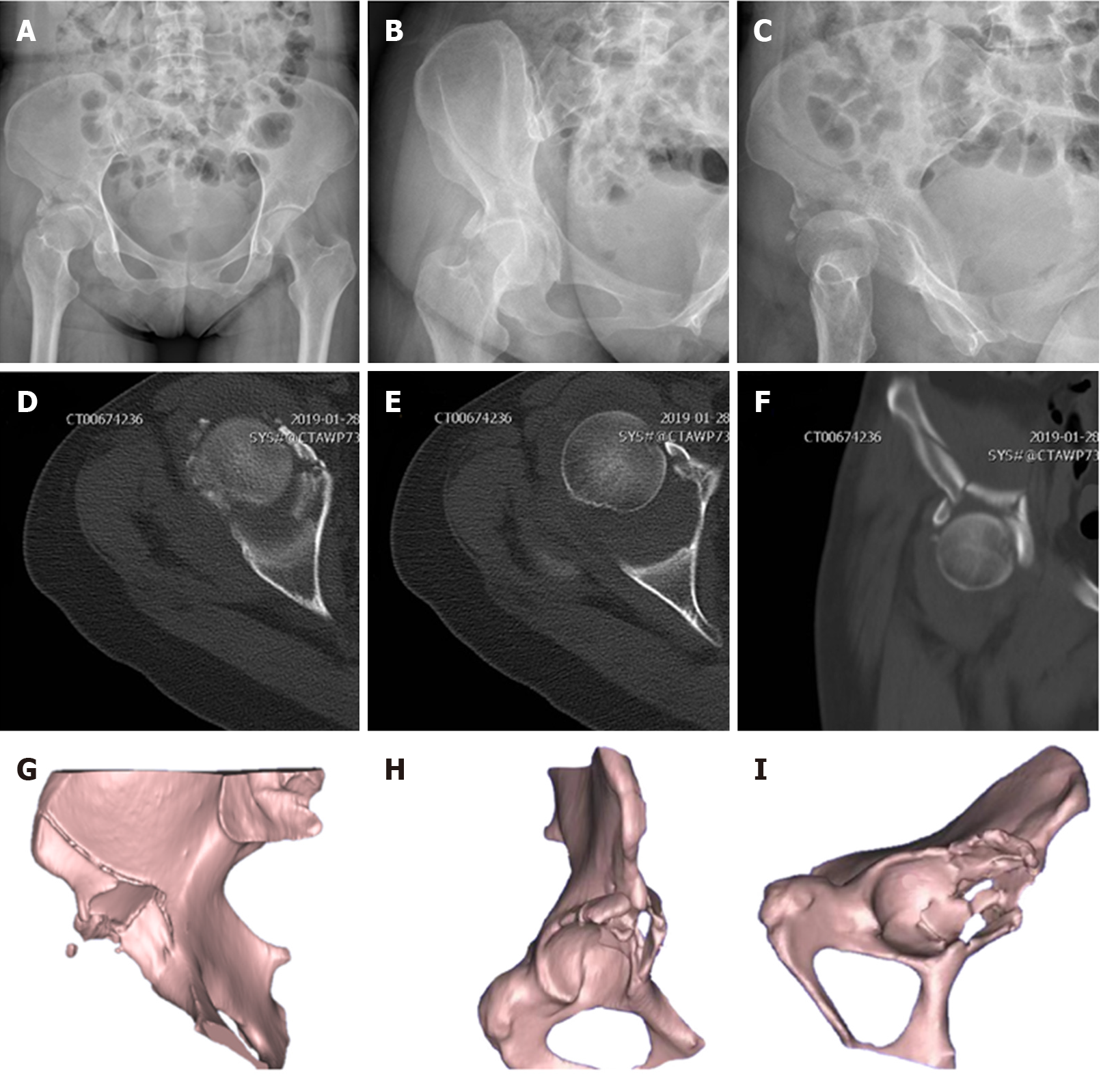
Figure 1 Preoperative plain radiographs and computed tomography scans.
A: Anteroposterior view radiography; B: Obturator oblique view radiography; C: Iliac oblique view radiography; D and E: Axial computed tomography (CT) images; F: Coronal CT image; G-I: Three-dimensional reconstructed CT images.
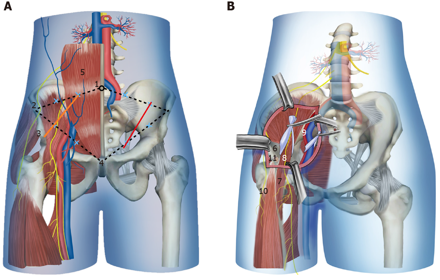
Figure 2 Schematic drawing demonstrating the skin incision and anatomic structures in the modified pararectus approach.
A: Skin incision; B: Anatomic structures. Orange line: Modified pararectus approach; Red line: Pararectus approach; 1: Umbilicus; 2: Anterior superior iliac spine; 3: Anterior inferior iliac spine; 4: Symphysis; 5: Rectus abdominis; 6: Iliac fossa; 7: Iliopsoas muscle; 8: Femoral nerve; 9: External iliac vessels; 10: Lateral femoral cutaneous nerve; 11: Avulsion fracture of anterior inferior iliac spine.
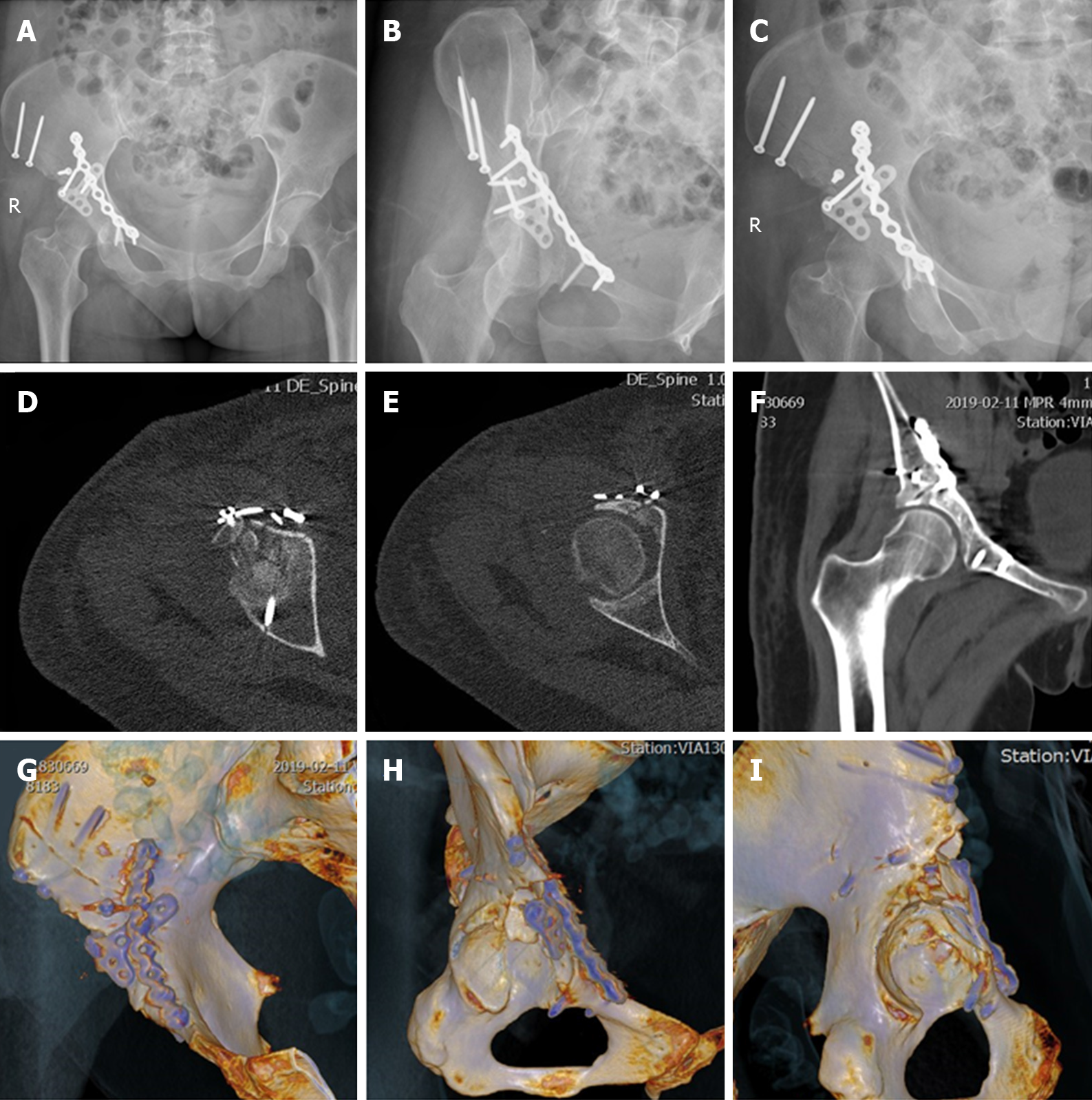
Figure 3 Postoperative plain radiographs and computed tomography scans.
A: Anteroposterior view radiography; B: Obturator oblique view radiography; C: Iliac oblique view radiography; D and E: Axial computed tomography (CT) images; F: Coronal CT image; G-I: Three-dimensional reconstructed CT images.
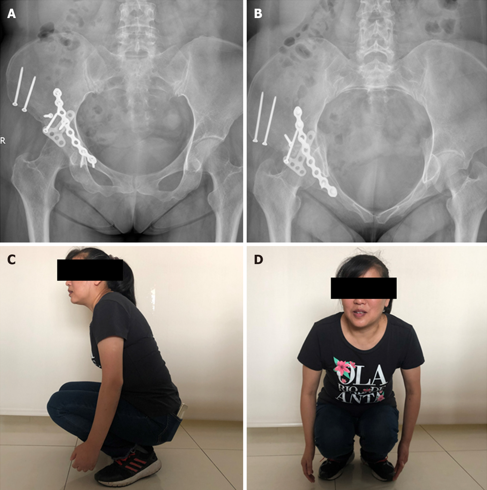
Figure 4 Postoperative X-rays and hip mobility at 9 mo.
A: Anteroposterior view radiography; B: Inlet view radiography; C and D: Flexion of the hip with a full load.
- Citation: Wang JJ, Ni JD, Song DY, Ding ML, Huang J, He GX, Li WZ. Modified pararectus approach for treatment of atypical acetabular anterior wall fracture: A case report. World J Clin Cases 2020; 8(12): 2634-2640
- URL: https://www.wjgnet.com/2307-8960/full/v8/i12/2634.htm
- DOI: https://dx.doi.org/10.12998/wjcc.v8.i12.2634


