Copyright
©The Author(s) 2020.
World J Clin Cases. Jun 26, 2020; 8(12): 2610-2616
Published online Jun 26, 2020. doi: 10.12998/wjcc.v8.i12.2610
Published online Jun 26, 2020. doi: 10.12998/wjcc.v8.i12.2610
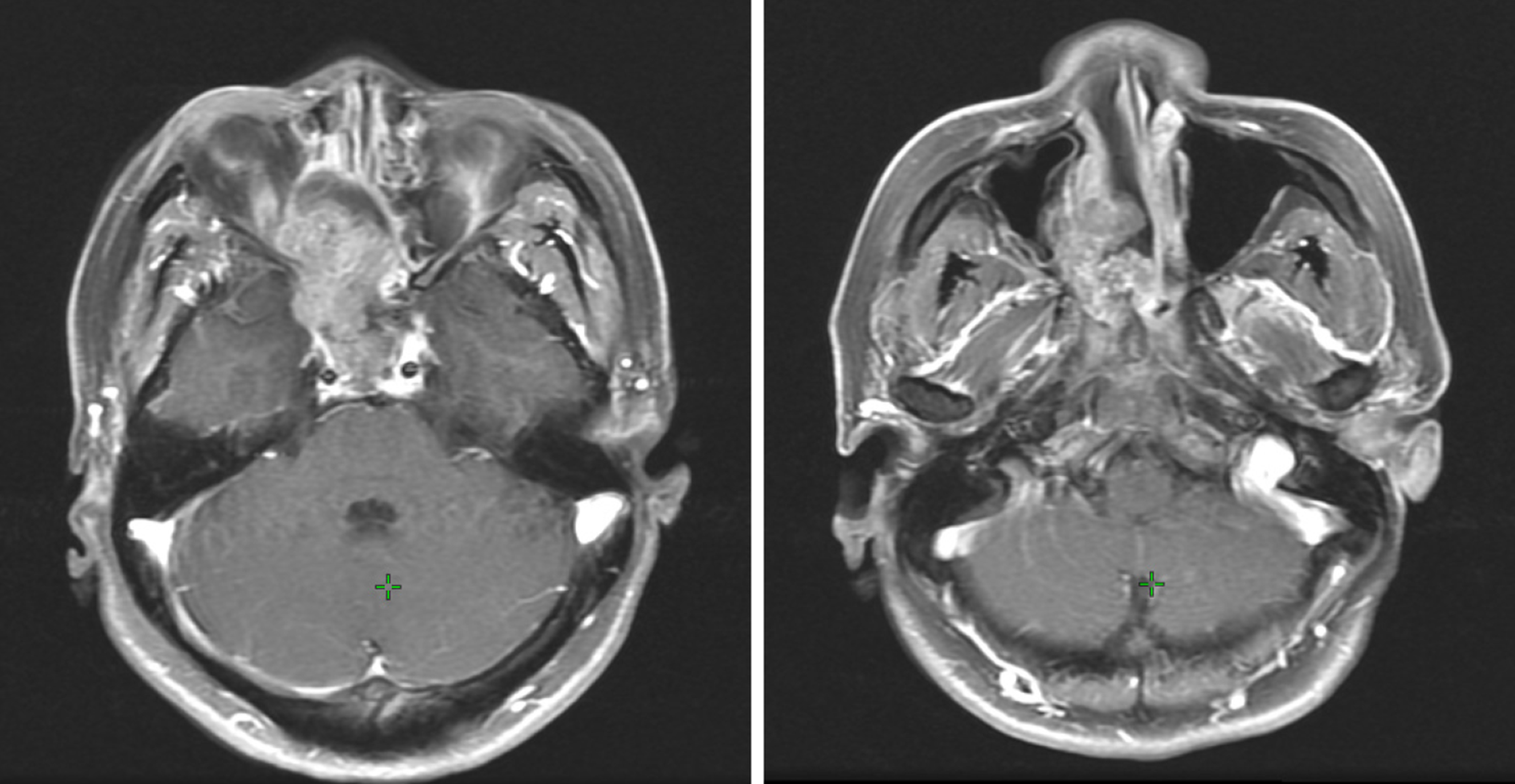
Figure 1 Images before surgery showing a mass in the nasal cavity and paranasal region.
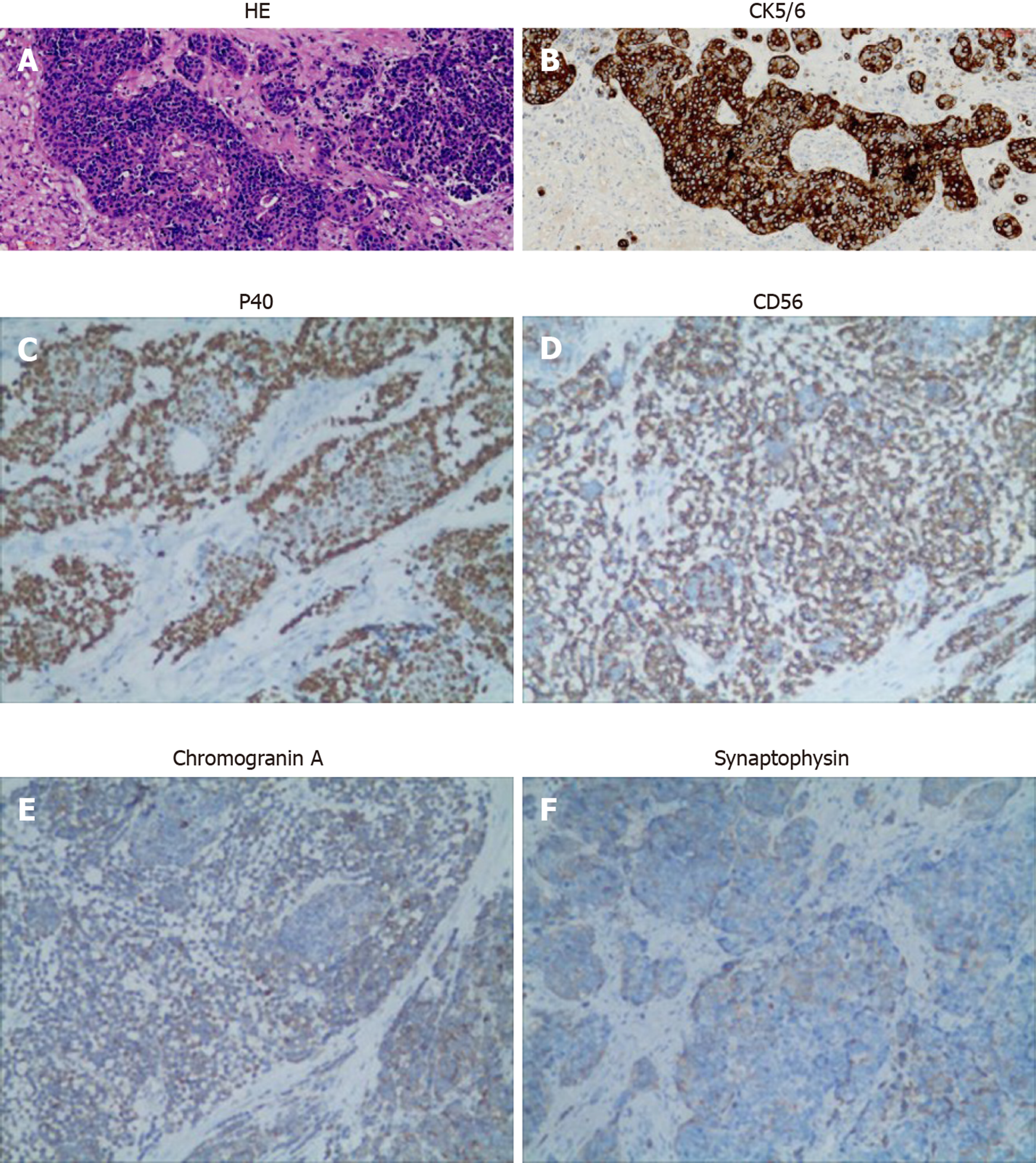
Figure 2 Immunohistochemical images.
A: Hematoxylin and eosin (HE) staining showed typical squamous and neuroendocrine differentiation areas of the neoplasm; B: CK5/6 was diffusely positive in the squamous differentiation area and negative in neuroendocrine differentiation cells; C: P40 was diffusely positive in the squamous differentiation area; D: CD56 was positive in neuroendocrine components; E: Chromogranin A expression was negative; F: Synaptophysin expression was negative. Original magnification, × 200.
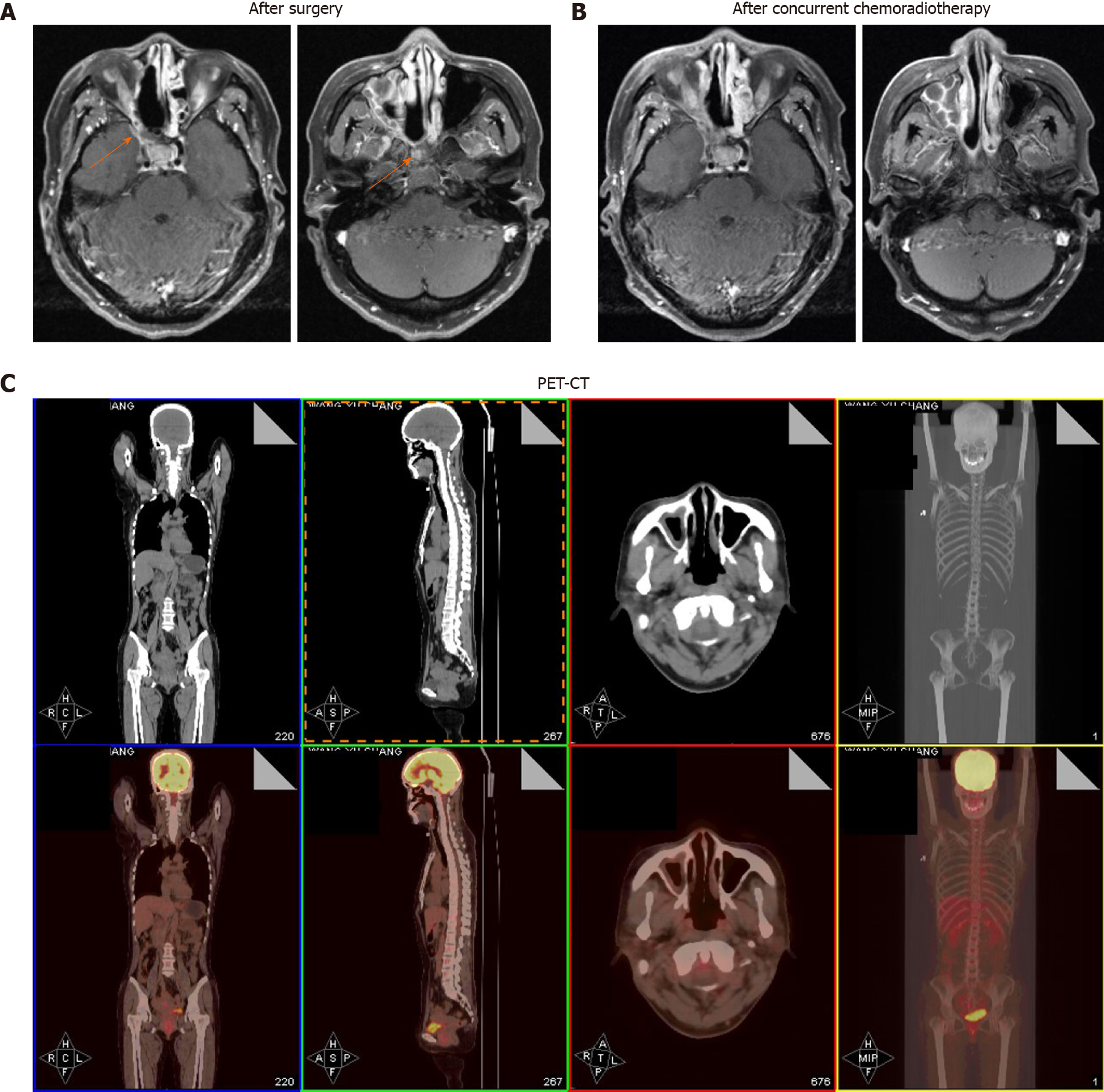
Figure 3 The patient has a complete response after chemoradiotherapy.
A: Images after surgery showing postoperative residual lesions (orange arrows); B: Images after surgery showing that a complete response was achieved; C: The latest positron emission tomography-computed tomography showing no signs of recurrence or metastasis.
- Citation: Wu SH, Zhang BZ, Han L. Collision tumor of squamous cell carcinoma and neuroendocrine carcinoma in the head and neck: A case report. World J Clin Cases 2020; 8(12): 2610-2616
- URL: https://www.wjgnet.com/2307-8960/full/v8/i12/2610.htm
- DOI: https://dx.doi.org/10.12998/wjcc.v8.i12.2610


