Copyright
©The Author(s) 2020.
World J Clin Cases. Jun 26, 2020; 8(12): 2585-2589
Published online Jun 26, 2020. doi: 10.12998/wjcc.v8.i12.2585
Published online Jun 26, 2020. doi: 10.12998/wjcc.v8.i12.2585
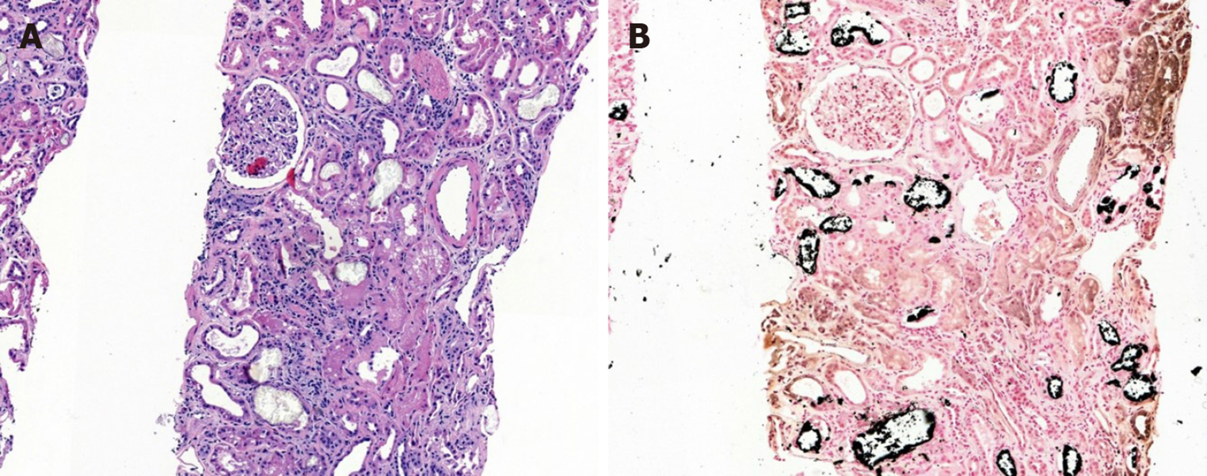
Figure 1 Renal biopsy specimen under light microscopy.
A: Hematoxylin and eosin stain; B: Von Kossa stain. Tubular calcium phosphate crystals staining positive. Magnification × 100.
- Citation: Medina-Liabres KRP, Kim BM, Kim S. Biopsy-proven acute phosphate nephropathy: A case report. World J Clin Cases 2020; 8(12): 2585-2589
- URL: https://www.wjgnet.com/2307-8960/full/v8/i12/2585.htm
- DOI: https://dx.doi.org/10.12998/wjcc.v8.i12.2585


