Copyright
©The Author(s) 2020.
World J Clin Cases. Jun 26, 2020; 8(12): 2530-2541
Published online Jun 26, 2020. doi: 10.12998/wjcc.v8.i12.2530
Published online Jun 26, 2020. doi: 10.12998/wjcc.v8.i12.2530
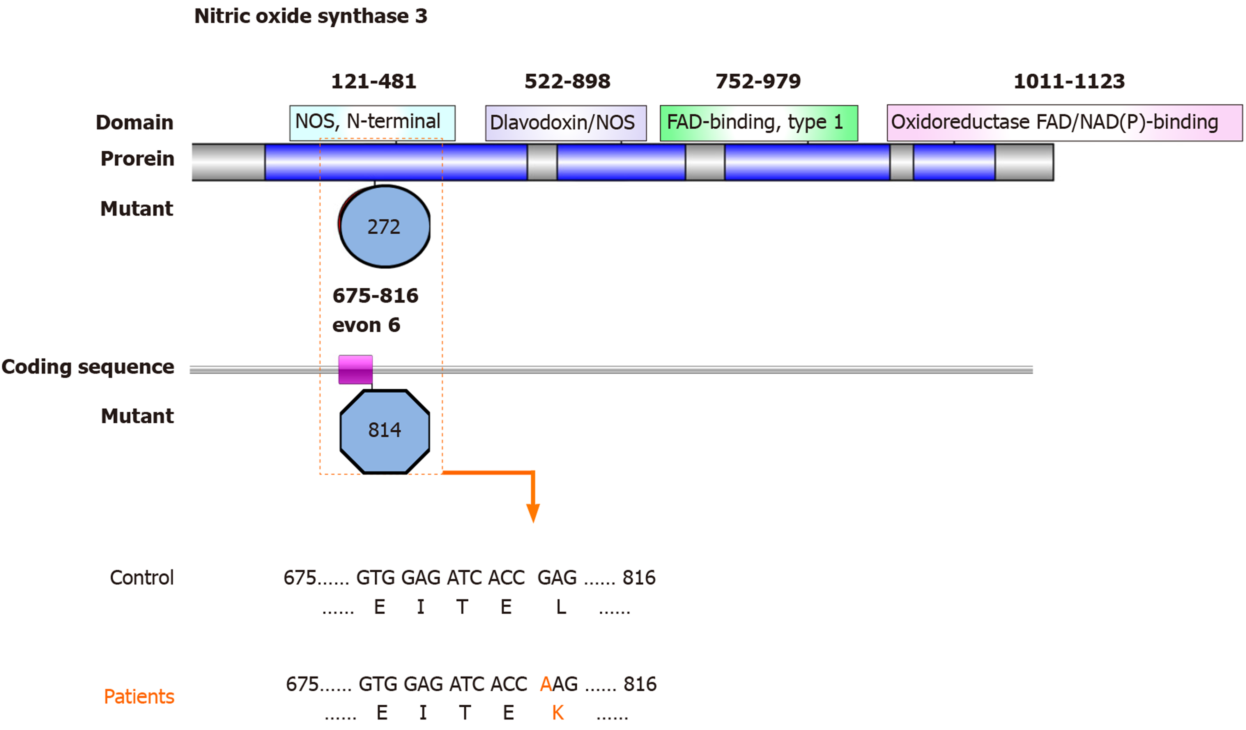
Figure 1 Map showing the nitric oxide synthase N-terminal domain with nitric oxide synthase 3 mutations identified in patients with osteonecrosis of the femoral head in systemic lupus erythematosus.
Top: Diagram of the nitric oxide synthase 3 (NOS3) protein structure. Nitric oxide synthase (NOS) comprises a NOS N-terminal domain (amino acids 121-481), a flavodoxin/NOS domain (amino acids 522-698), a flavin adenine dinucleotide-binding, type 1 domain (amino acids 752-979), and an oxidoreductase flavin adenine dinucleotide/nicotinamide adenine dinucleotide (P)-binding domain (amino acids 1011-1123). Middle: The novel mutation of the NOS3 gene coding sequence identified in this study, located in exon 6 (encoding amino acids 675-816). Bottom: Mutated nucleotides in exon 6 of NOS3 are shown in orange. Mutated amino acids in the NOS N-terminal domain of NOS3 are shown in orange. FAD-binding: Flavin adenine dinucleotide-binding; NAD: Nicotinamide adenine dinucleotide.
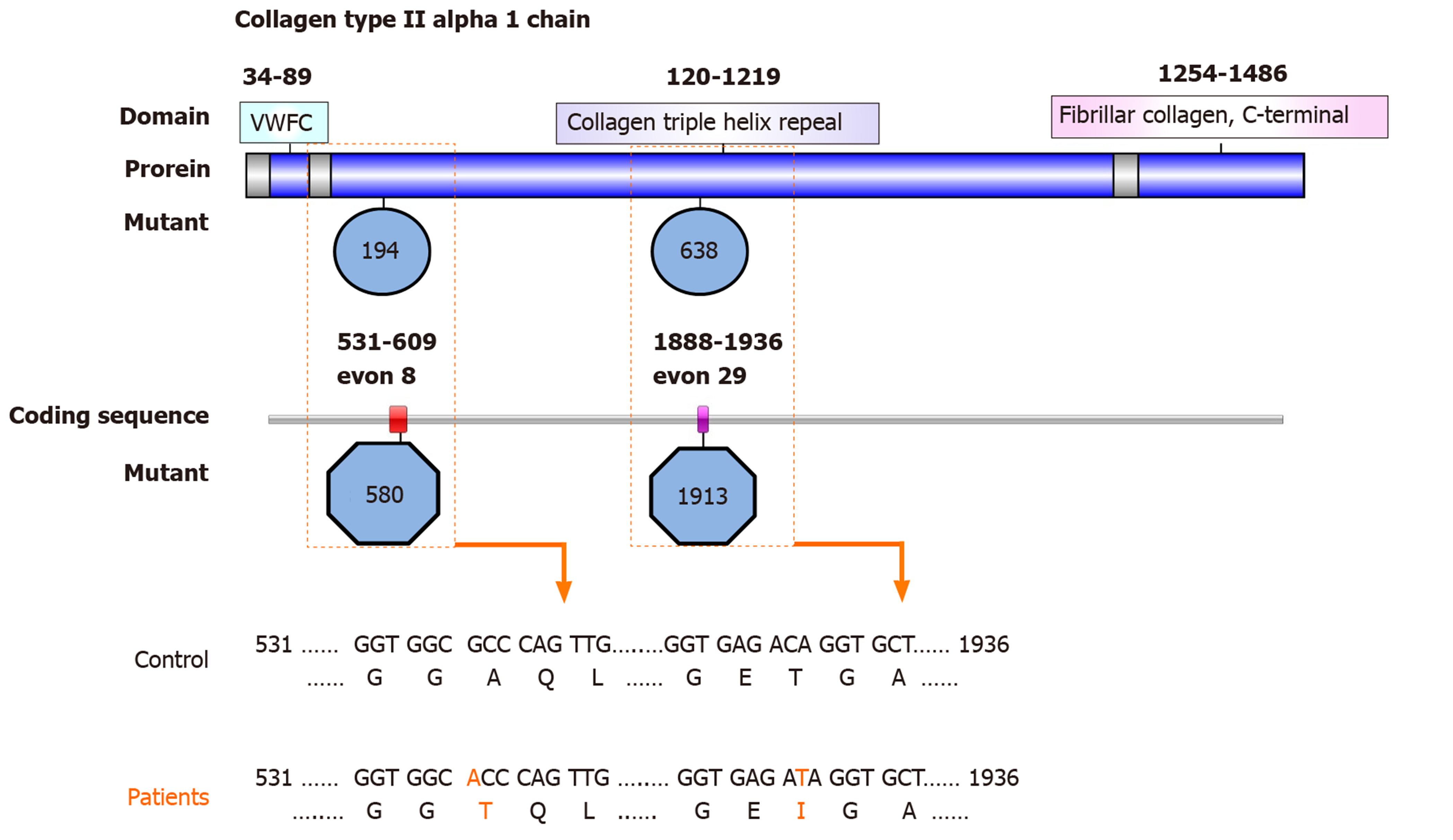
Figure 2 Map showing the collagen triple helix repeat domain with collagen type II alpha 1 chain mutations identified in patients with osteonecrosis of the femoral head in systemic lupus erythematosus.
Top: Diagram of the collagen type II alpha 1 chain (COL2A1) protein structure. The COL2A1 comprises a von Willebrand Factor C (VWFC) domain (amino acids 34-89), a collagen triple helix repeat domain (amino acids 120-1219), and a fibrillar collagen C-terminal domain (amino acids 1254-1486). Middle: The novel mutations identified in the COL2A1 gene coding sequence in this study are located in exon 8 (encoding amino acids 531-609) and exon 29 (encoding amino acids 1888-1936). Bottom: Mutated nucleotides in exons 8 and 29 of the COL2A1 gene are shown in orange. Mutated amino acids in the VWFC and the collagen triple helix repeat domains of COL2A1 are shown in orange.
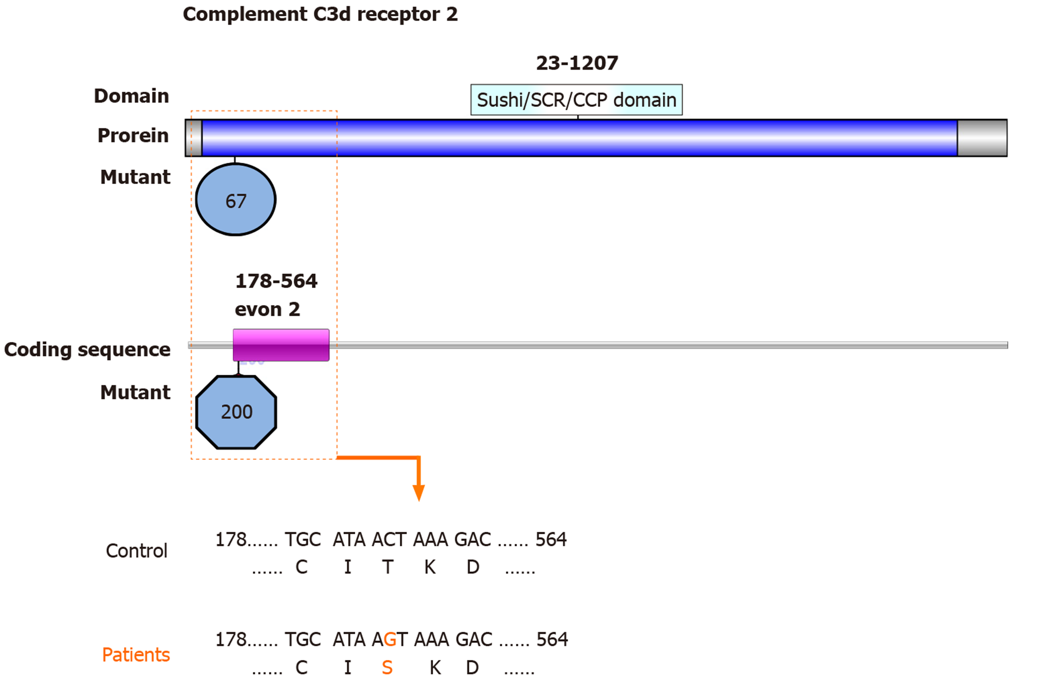
Figure 3 Map of the Sushi/short consensus repeat/complement control protein domain of complement C3d receptor 2 with mutations identified in patients with osteonecrosis of the femoral head in systemic lupus erythematosus.
Top: Diagram of the complement C3d receptor 2 (CR2) protein structure. CR2 comprises a Sushi/short consensus repeat/complement control protein domain (amino acids 23-1027). Middle: The novel mutation identified in the CR2 gene coding sequence in this study is located in exon 2 (encoding amino acids 178-564) Bottom: Mutated nucleotides in exon 2 of CR2 are shown in orange. Mutated amino acids in the Sushi/short consensus repeat/complement control protein domain of CR2 are shown in orange. CCP: Complement control protein; SCR: Short consensus repeat.
- Citation: Sun HS, Yang QR, Bai YY, Hu NW, Liu DX, Qin CY. Gene testing for osteonecrosis of the femoral head in systemic lupus erythematosus using targeted next-generation sequencing: A pilot study. World J Clin Cases 2020; 8(12): 2530-2541
- URL: https://www.wjgnet.com/2307-8960/full/v8/i12/2530.htm
- DOI: https://dx.doi.org/10.12998/wjcc.v8.i12.2530


