Copyright
©The Author(s) 2020.
World J Clin Cases. Jun 26, 2020; 8(12): 2502-2509
Published online Jun 26, 2020. doi: 10.12998/wjcc.v8.i12.2502
Published online Jun 26, 2020. doi: 10.12998/wjcc.v8.i12.2502
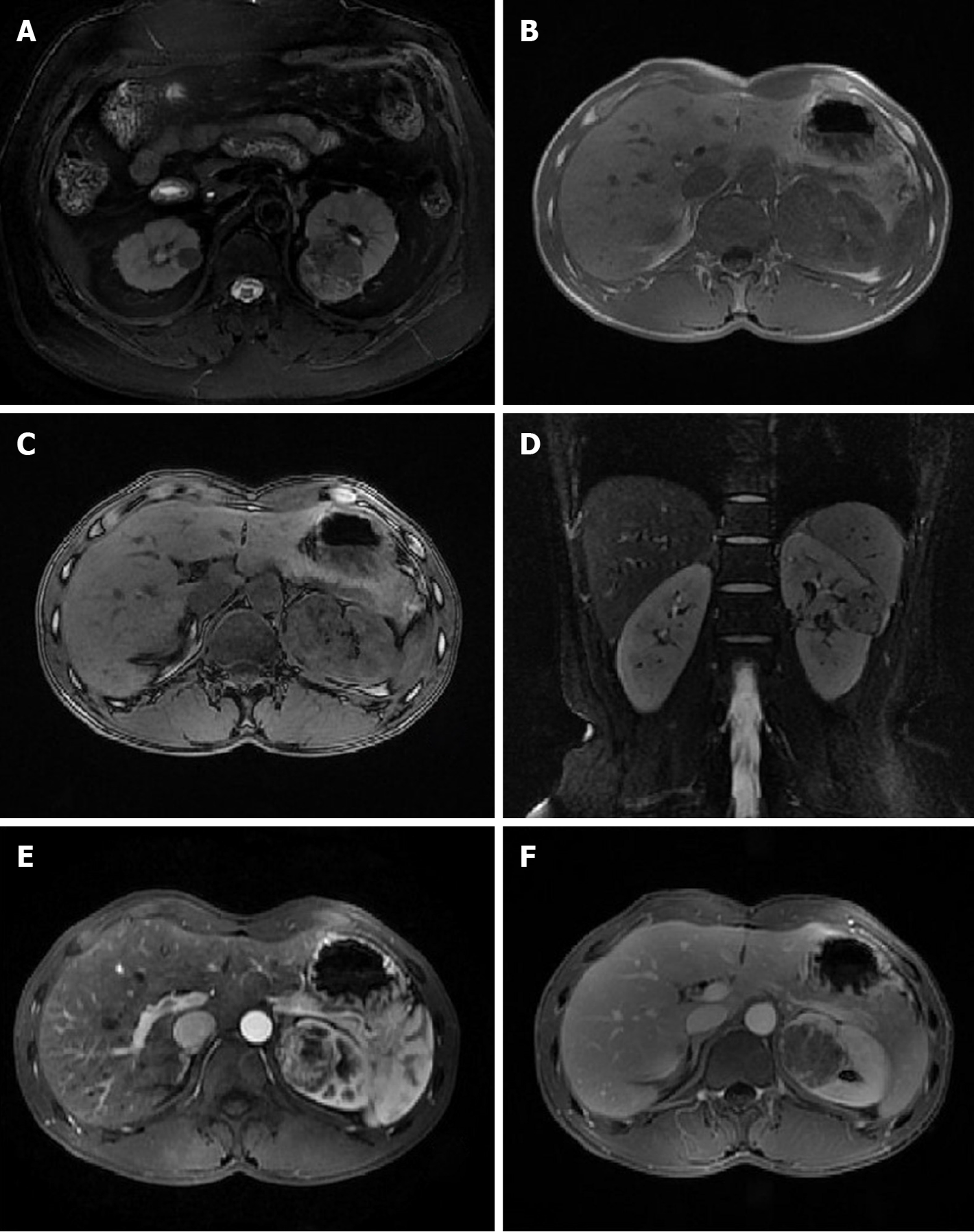
Figure 1 Intratumoral vasculature was not observed in eight cases but was visible in the other two.
A: An oval, homogeneous, and short T2 signal was found in the right kidney, showing a surrounding capsule. The lesion in the left kidney was pathologically confirmed as renal clear cell carcinoma; B and C: A mass in the renal parenchyma of the left kidney showed decreased signal intensity on opposed-phase MRI, indicating lipid composition; D: An oval, short T2 signal was found in the left kidney, showing a surrounding capsule; E and F: The round-like tumor in the left kidney showed heterogeneous and significant enhancement in cortico-medullary phase and decreased signal intensity(washout) of tumor in the excretory phase.
- Citation: Li XL, Shi LX, Du QC, Wang W, Shao LW, Wang YW. Magnetic resonance imaging features of minimal-fat angiomyolipoma and causes of preoperative misdiagnosis. World J Clin Cases 2020; 8(12): 2502-2509
- URL: https://www.wjgnet.com/2307-8960/full/v8/i12/2502.htm
- DOI: https://dx.doi.org/10.12998/wjcc.v8.i12.2502


