Copyright
©The Author(s) 2020.
World J Clin Cases. Jun 26, 2020; 8(12): 2408-2424
Published online Jun 26, 2020. doi: 10.12998/wjcc.v8.i12.2408
Published online Jun 26, 2020. doi: 10.12998/wjcc.v8.i12.2408
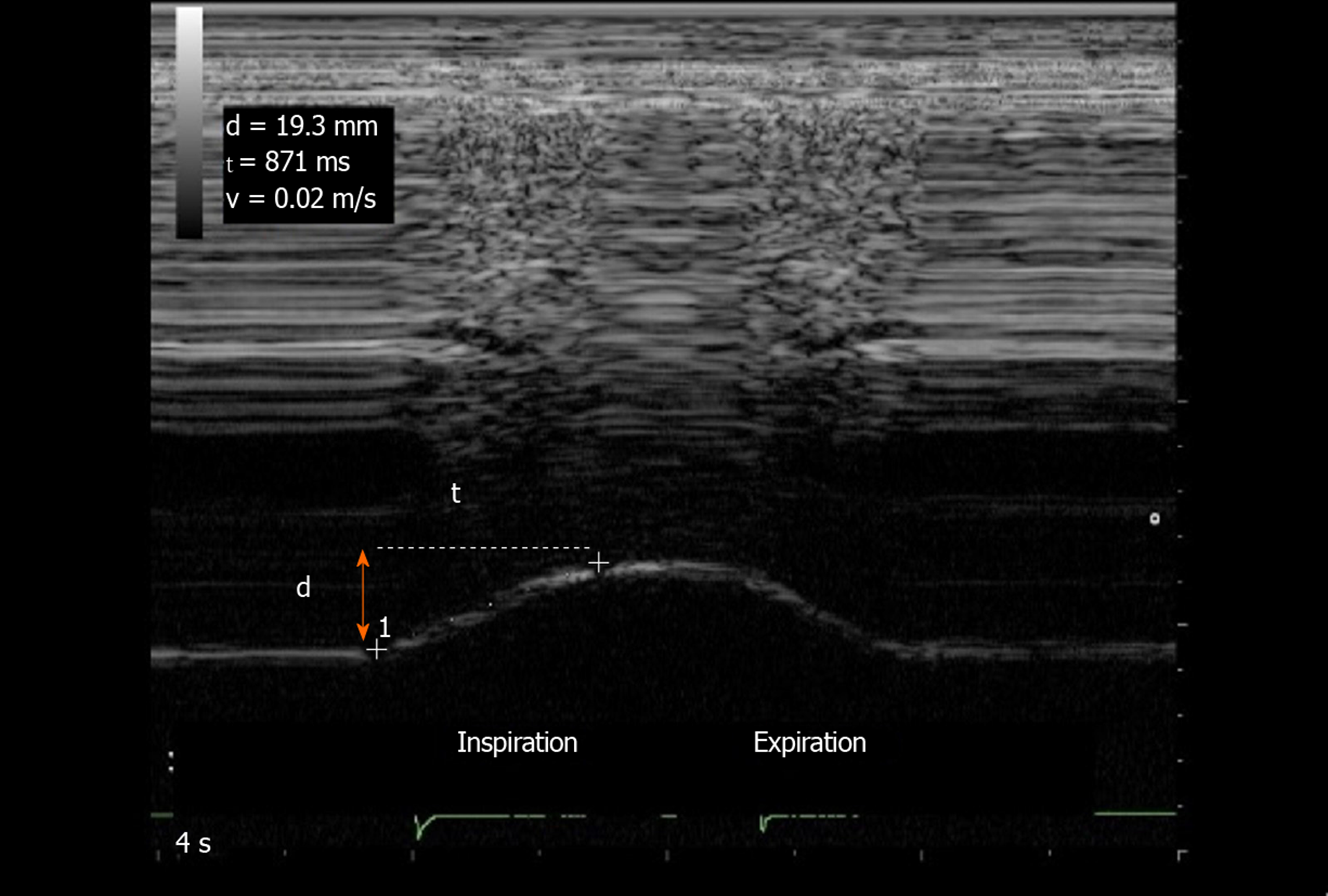
Figure 1 Diaphragmatic motion recorded by M-mode ultrasonography: measurement of diaphragm excursion, inspiratory time and velocity of contraction.
Ordinate in centimeter, abscissa in second. d: Diaphragm excursion; t: Inspiratory time; v: Velocity of contraction.
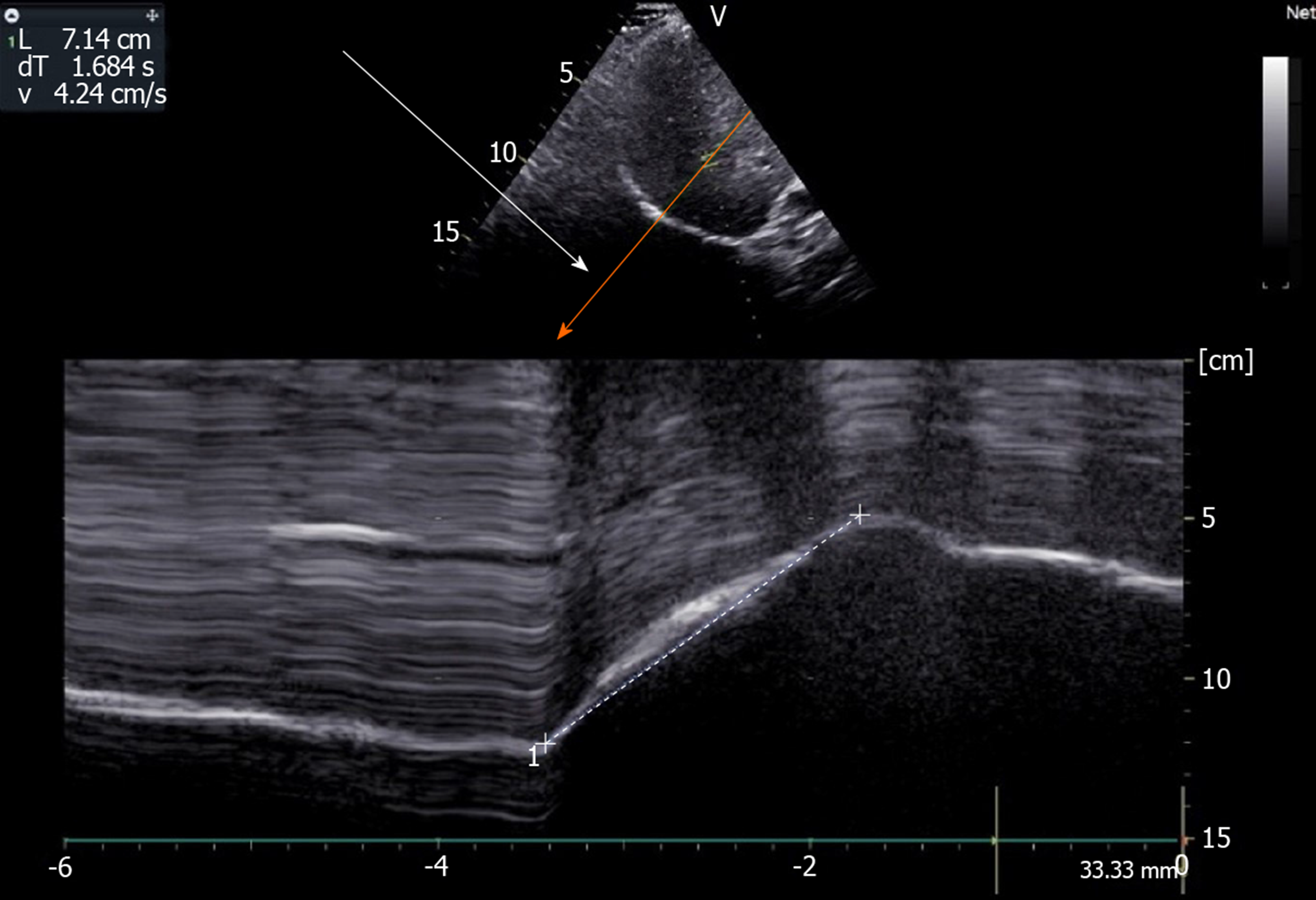
Figure 2 Use of angle-independent M-mode sonography (arrow) to obtain a perpendicular approach of the right hemidiaphragm: Measurement of excursion (7.
14 cm) during deep breathing. Ordinate in centimeter, abscissa in second.
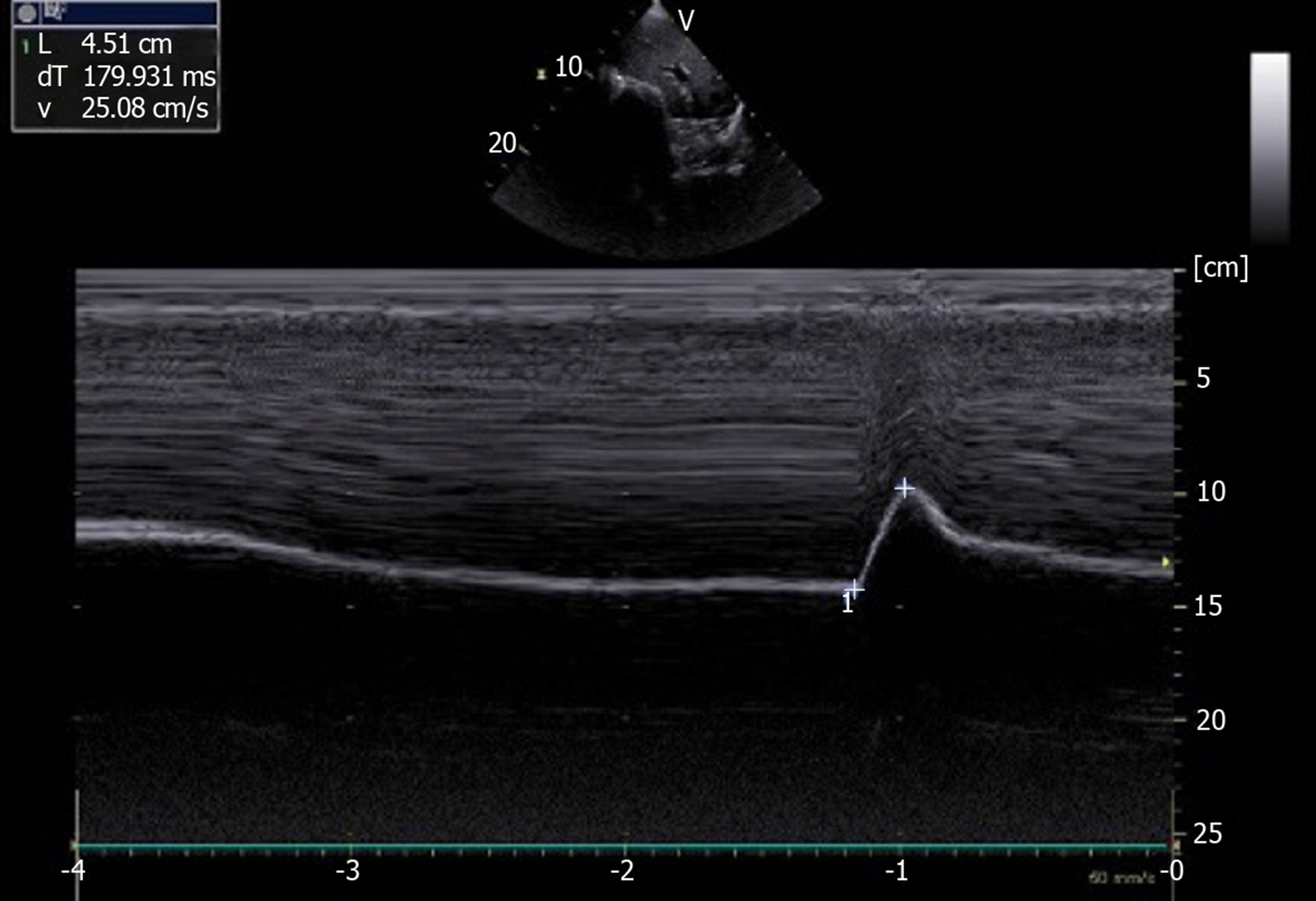
Figure 3 Diaphragmatic motion recorded by M-mode ultrasonography during voluntary sniffing (excursion = 4.
51 cm). Ordinate in centimeter, abscissa in second.
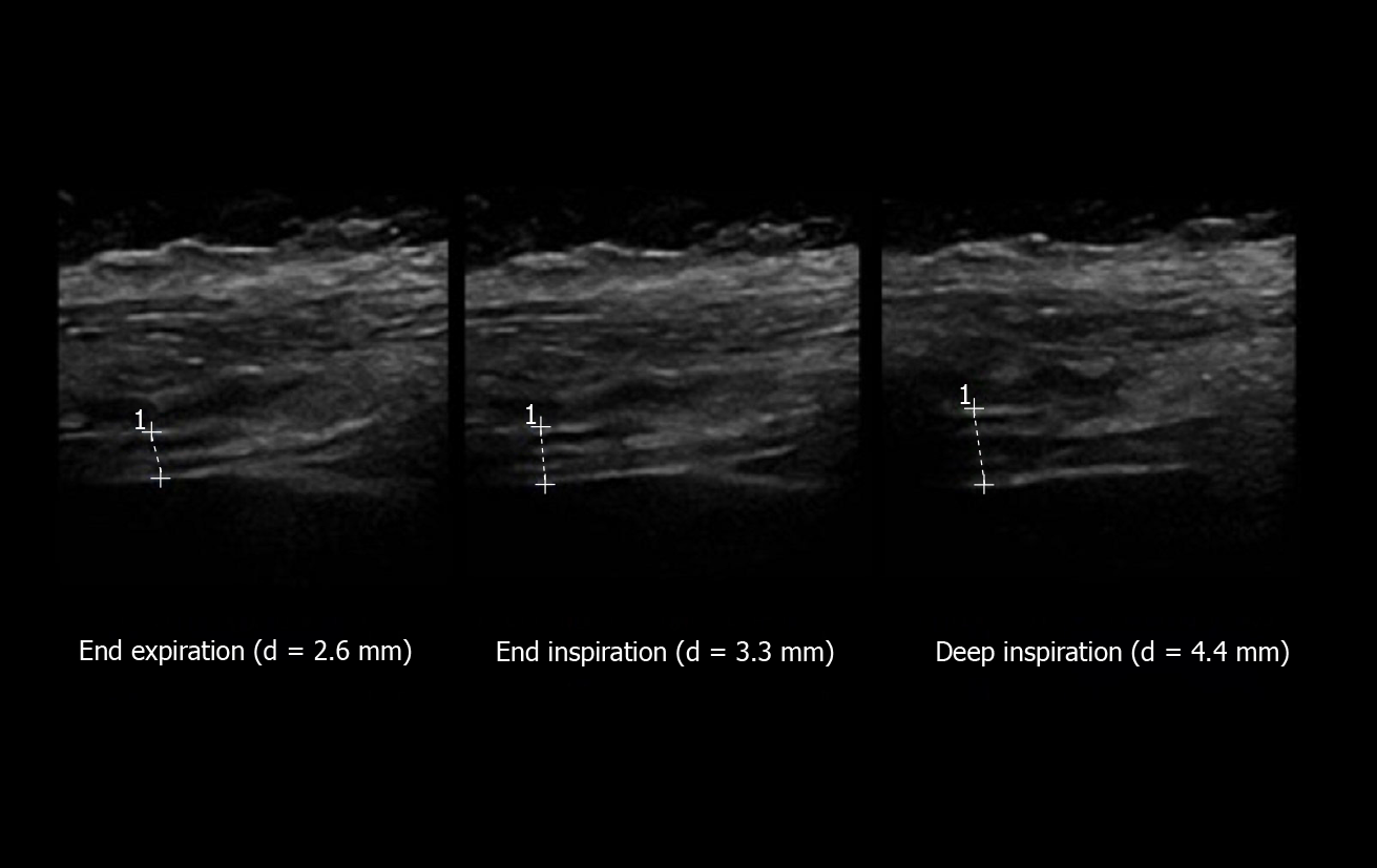
Figure 4 Measurement of diaphragm thickness using B-mode ultrasonography: The diaphragm thickening is calculated from the measurement of thickness at both end expiration and end inspiration [here = (4.
4-2.6)/2.6 = 69%]. Ordinate in centimeter, abscissa in second.
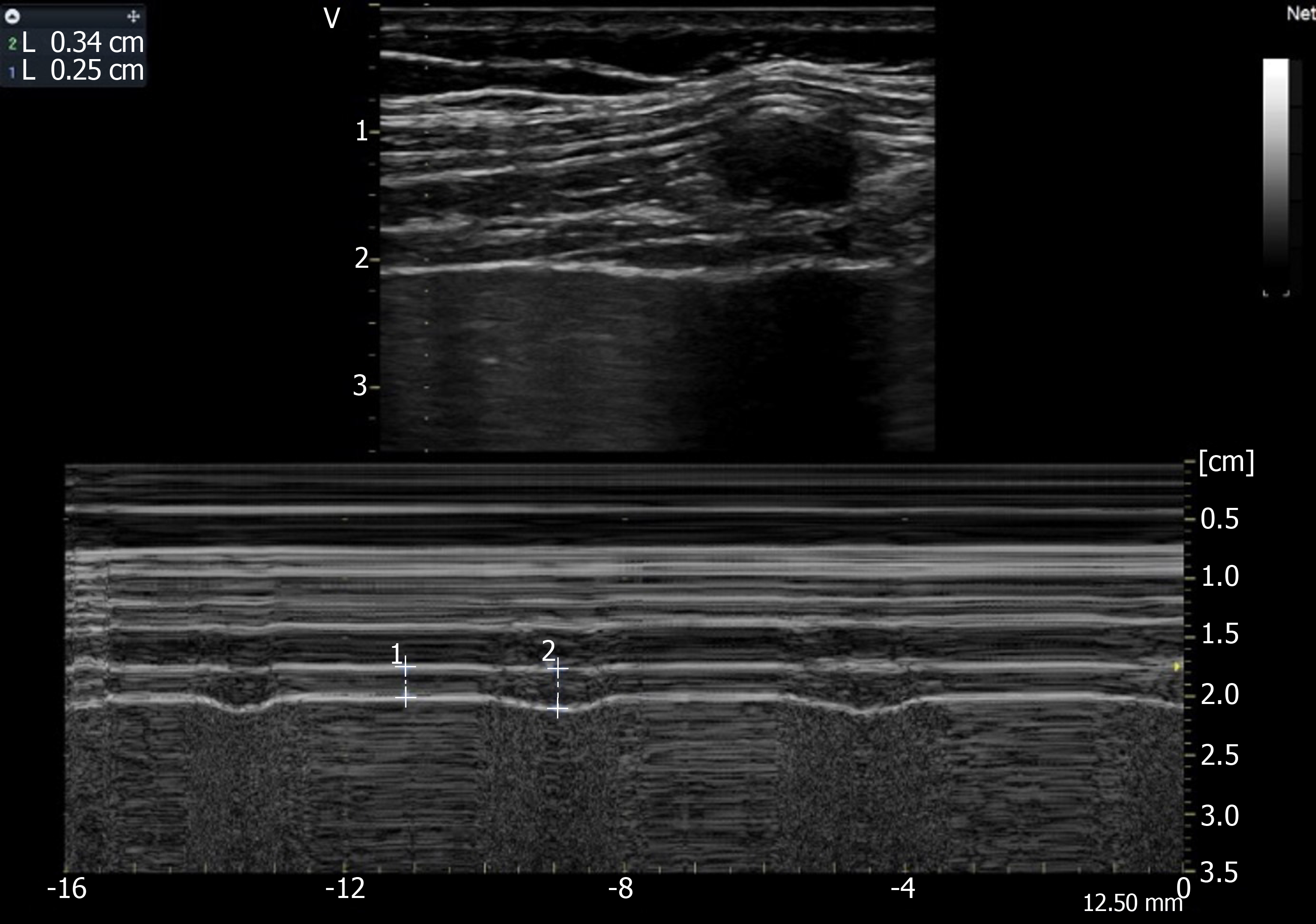
Figure 5 Recording of the changes in diaphragm thickness during quiet breathing using M-mode tracing (measurement of thickness at end expiration (1 L = 0.
25 cm), at end inspiration (2 L = 0.34 cm). Ordinate in centimeter, abscissa in second.
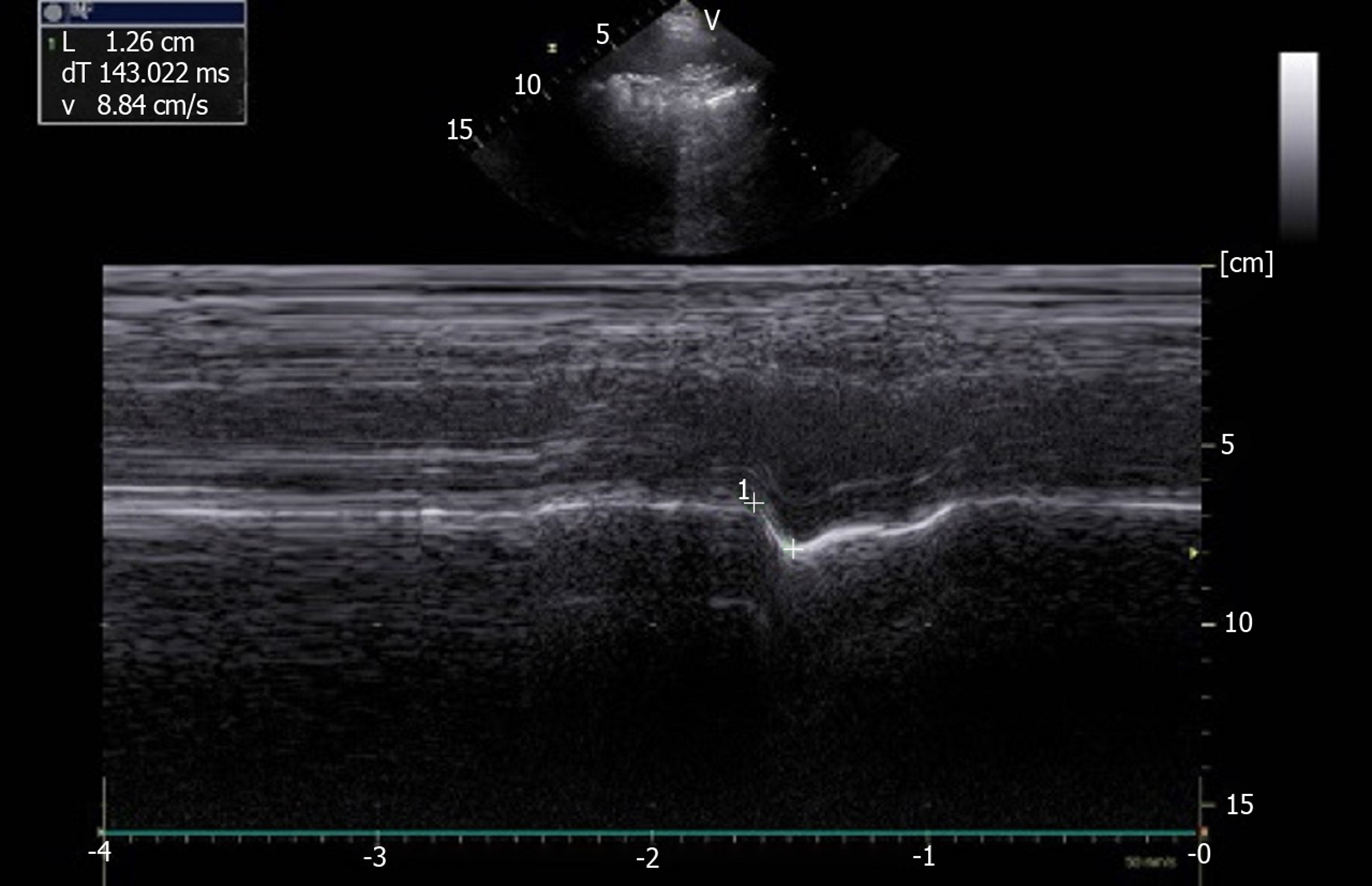
Figure 6 Paradoxical motion (-1.
26 cm) recorded by M-mode ultrasonography in patient with left hemidiaphragm paralysis. Ordinate in centimeter, abscissa in second.
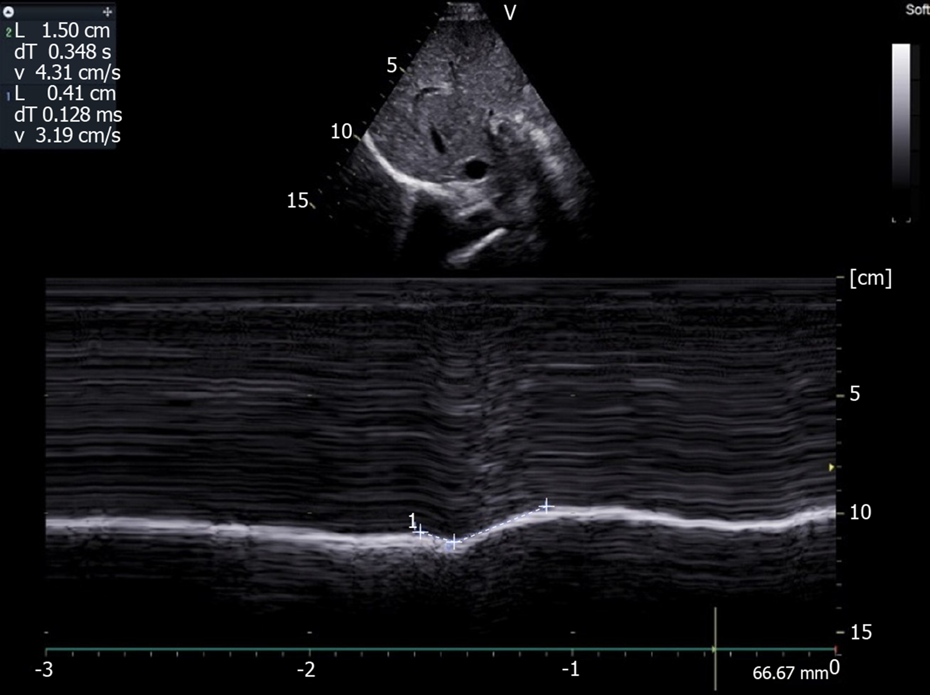
Figure 7 Motion recorded during deep breathing in patient suffering from right hemidiaphragm paralysis: Paradoxical motion at the beginning of inspiration (-0.
41 cm) terminal excursion in the cranio-caudal direction (+1.5 cm). Ordinate in centimeter, abscissa in second.
- Citation: Boussuges A, Rives S, Finance J, Brégeon F. Assessment of diaphragmatic function by ultrasonography: Current approach and perspectives. World J Clin Cases 2020; 8(12): 2408-2424
- URL: https://www.wjgnet.com/2307-8960/full/v8/i12/2408.htm
- DOI: https://dx.doi.org/10.12998/wjcc.v8.i12.2408


