Copyright
©The Author(s) 2020.
World J Clin Cases. Jun 6, 2020; 8(11): 2318-2324
Published online Jun 6, 2020. doi: 10.12998/wjcc.v8.i11.2318
Published online Jun 6, 2020. doi: 10.12998/wjcc.v8.i11.2318
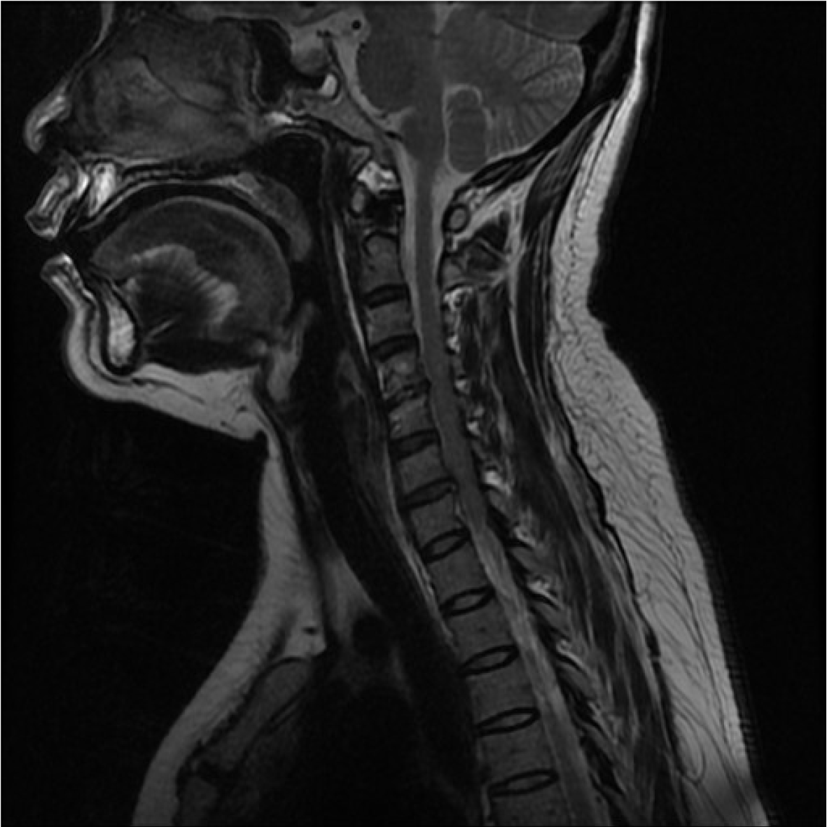
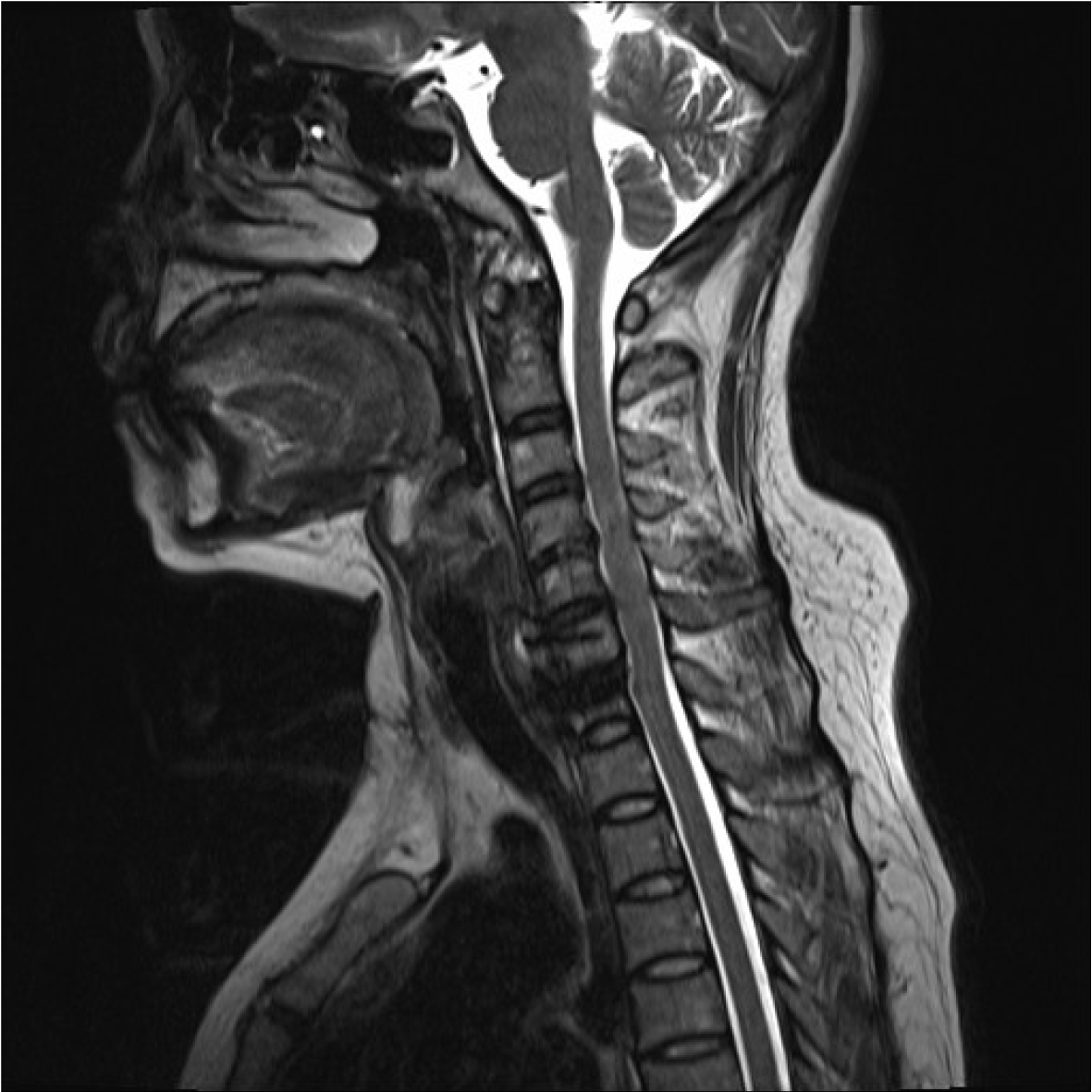
Figure 1 Magnetic resonance imaging showed obvious degeneration of C4/5 disc with Modic type 2 changes, and slight herniation of C6/7 disc without compression of the spinal cord or nerve root.
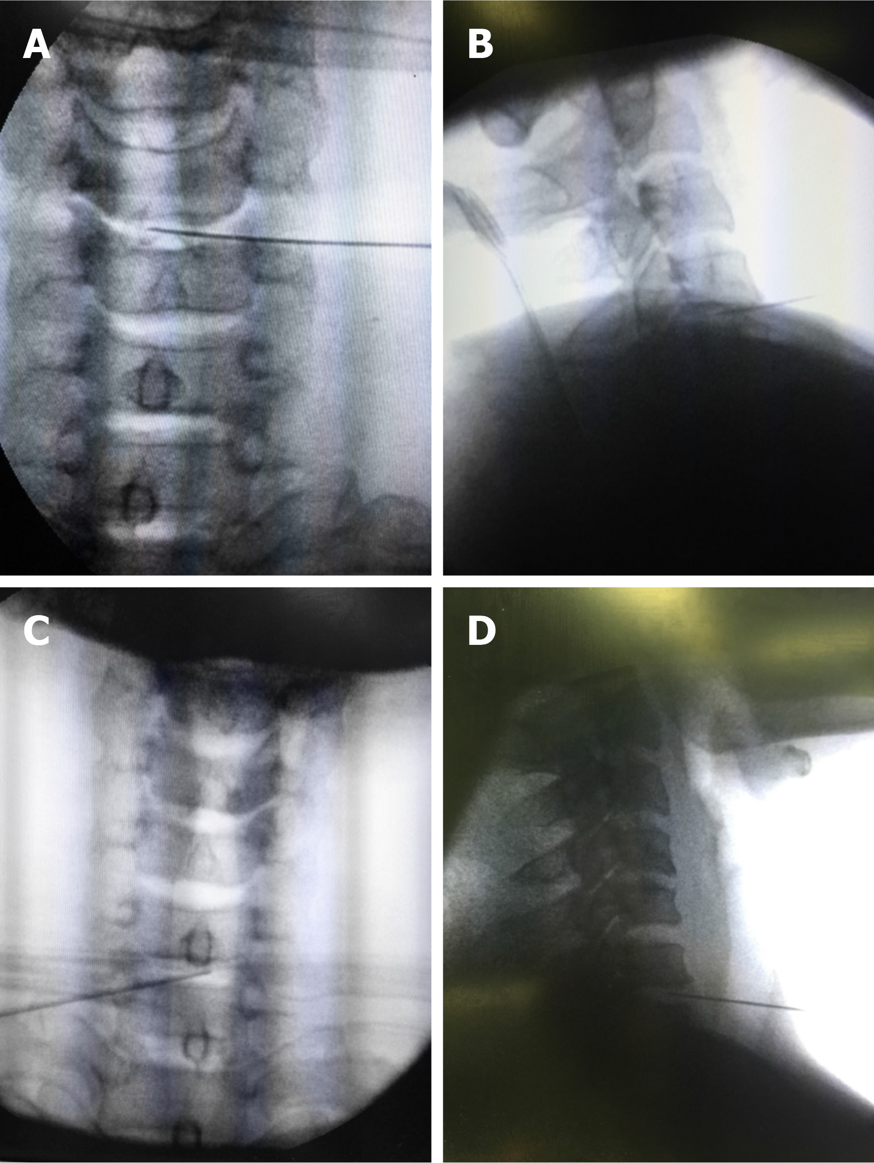
Figure 2 Image of stages in cervical intradiscal analgesia.
A: Anterior view of a needle inserted into C4/5 intervertebral disc; B: Lateral view of the needle inserted; C: Anterior view of a needle inserted into C6/7 intervertebral disc; D: Lateral view of the needle inserted.
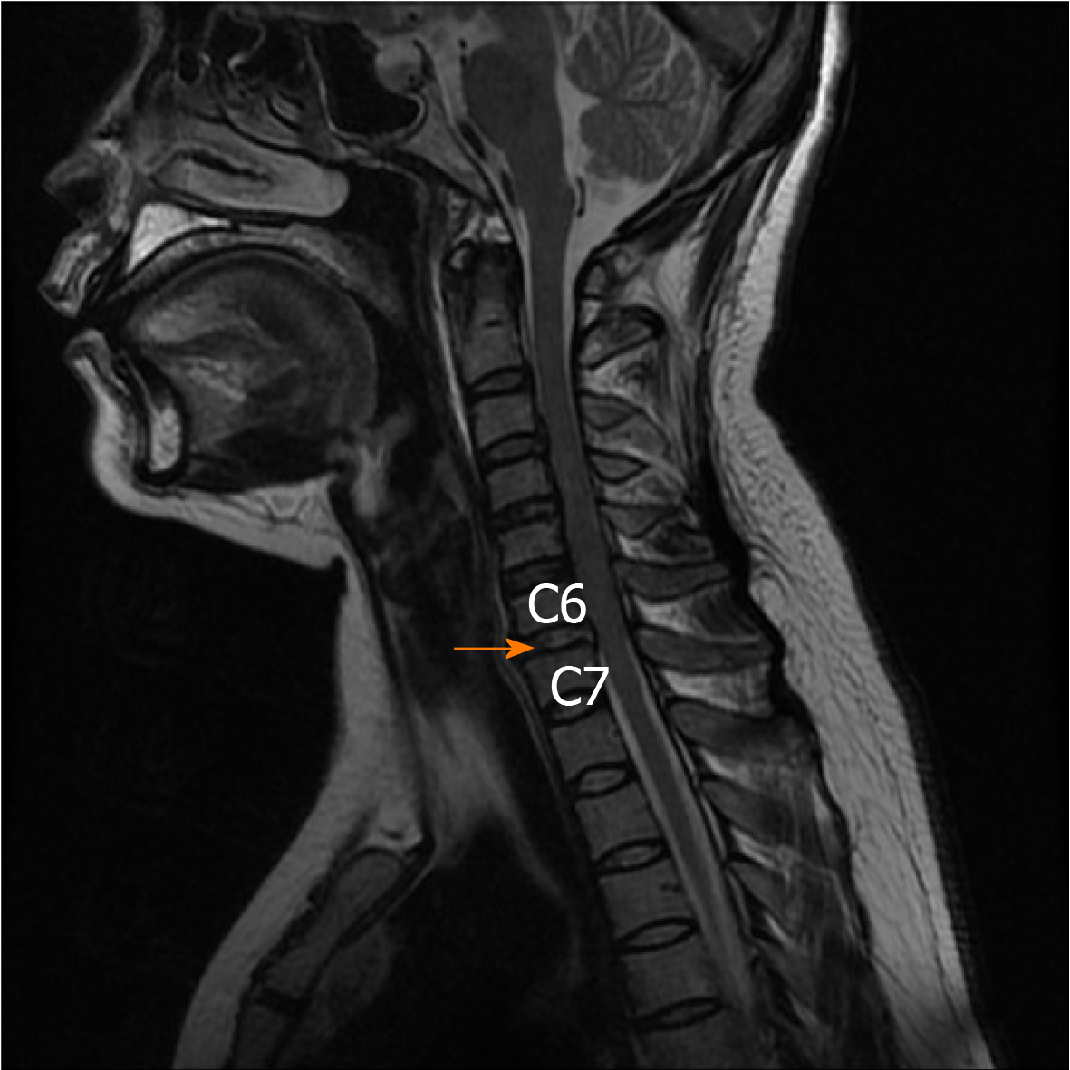
Figure 3 At 5 d after the second injection, the cervical 6/7 disc (arrow) showed increased signal intensity.
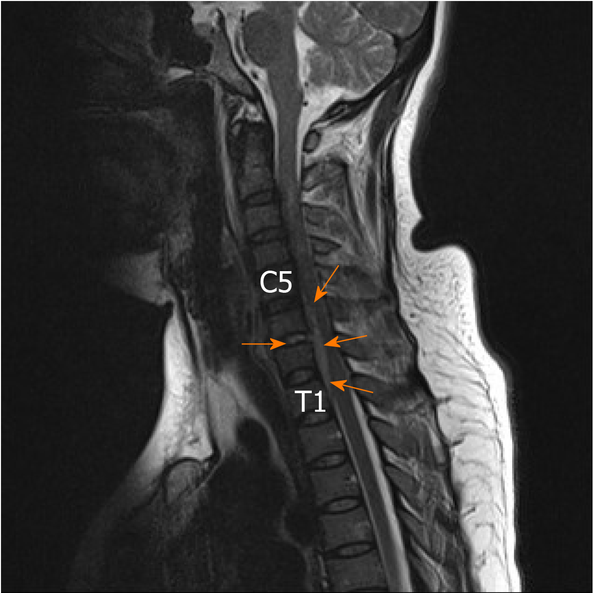
Figure 4 At 10 d after the second injection, epidural inflammation (arrows) could be seen with increased signal intensity from the lower margin of C5 to the lower margin of T1 as well as within the C6/7 disc.
Figure 5 Magnetic resonance imaging showed that the epidural abscess was almost completely absorbed, and the epidural space showed normal signal intensity.
- Citation: Wu B, He X, Peng BG. Pyogenic discitis with an epidural abscess after cervical analgesic discography: A case report. World J Clin Cases 2020; 8(11): 2318-2324
- URL: https://www.wjgnet.com/2307-8960/full/v8/i11/2318.htm
- DOI: https://dx.doi.org/10.12998/wjcc.v8.i11.2318


