Copyright
©The Author(s) 2020.
World J Clin Cases. May 26, 2020; 8(10): 1932-1938
Published online May 26, 2020. doi: 10.12998/wjcc.v8.i10.1932
Published online May 26, 2020. doi: 10.12998/wjcc.v8.i10.1932
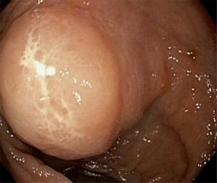
Figure 1 Findings at colonoscopy.
A 3 cm submucosal pedunculated polyp is seen about 15 cm from anal verge.
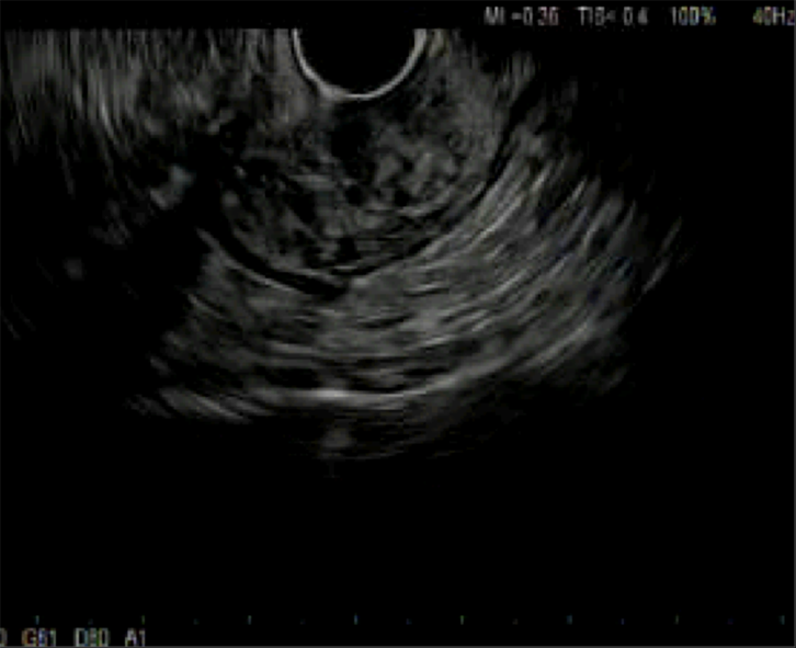
Figure 2 Findings with endoscopic ultrasound.
A 2.8 cm × 15.2 cm avascular lesion of mixed echogenicity seen in the submucosa with no communication with muscularis mucosa or propria.
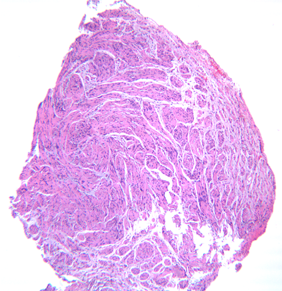
Figure 3 Histopathology of the tumor at low power × 10 magnification.
Bland spindle cell tumor is seen in a fascicular growth pattern.
Figure 4 Immunohistochemistry of the lesion at × 50 magnification.
Tumor is diffusely positive for S100. Note the penetrating axons with neurofilament stain.
- Citation: Ghoneim S, Sandhu S, Sandhu D. Isolated colonic neurofibroma, a rare tumor: A case report and review of literature. World J Clin Cases 2020; 8(10): 1932-1938
- URL: https://www.wjgnet.com/2307-8960/full/v8/i10/1932.htm
- DOI: https://dx.doi.org/10.12998/wjcc.v8.i10.1932


