Copyright
©The Author(s) 2020.
World J Clin Cases. Jan 6, 2020; 8(1): 126-132
Published online Jan 6, 2020. doi: 10.12998/wjcc.v8.i1.126
Published online Jan 6, 2020. doi: 10.12998/wjcc.v8.i1.126
Figure 1 The tumor and orthopantomography.
A: A tumor mass affecting the palatal and vestibular regions of the left maxilla was observed. The palatal mucosa was normochromic and had a retained canine. In the vestibular sulcus, there was multilobulated tumor mass with a yellowish and telangiectatic surface; B: Orthopantomography showing a poorly defined radiolucent zone apparently associated with a canine retained at the level of the left upper premolars.
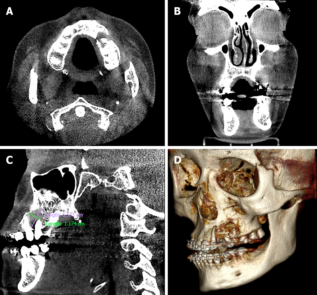
Figure 2 Computed tomography with 3-dimensional reconstruction.
A, B: Computed axial tomograph; C, D: Affected vestibular and palatal and cortical bones. Size, delimitation, and degree of destruction of affected bone tissue.
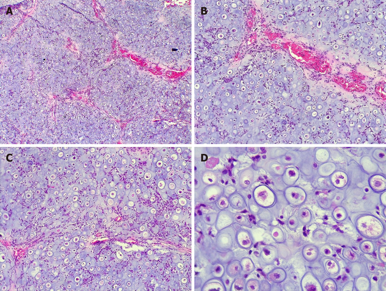
Figure 3 Hematoxylin and eosin stain.
A: 50 ×; B: 100 ×; Cartilaginous neoplasia with well-vascularized lobular pattern; C: 100 ×; D: 400 ×. Proliferation of hyperchromatic, binucleated, and pleomorphic chondrocytes in large and irregular lagoons.
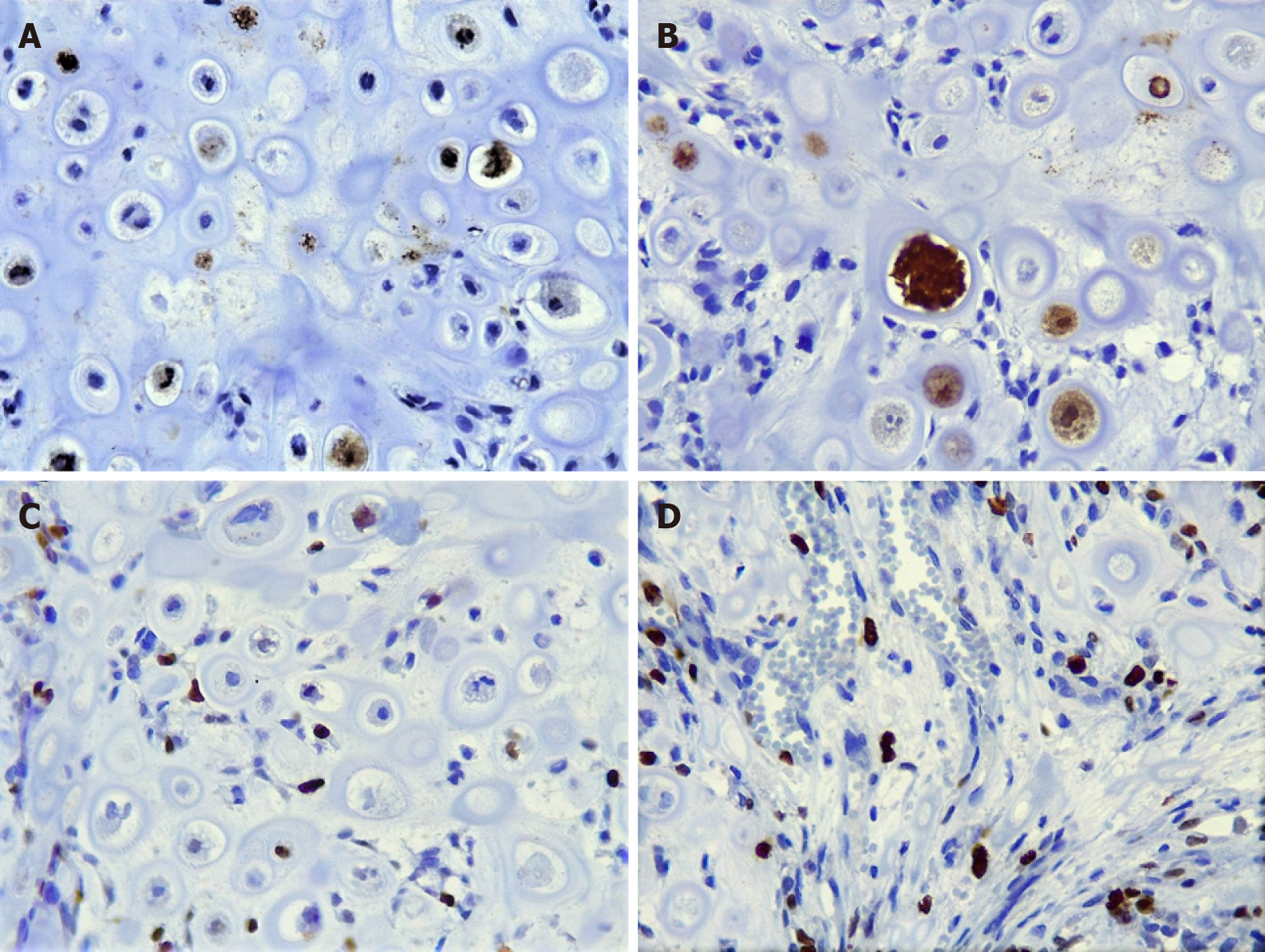
Figure 4 Hematoxylin and eosin stain.
A, B: Pleomorphic chondrocytes with nuclear positivity for S-100 (400 ×); C, D: Moderate reaction to Ki-67 (400 ×); proliferative activity is observed mainly in undifferentiated small cells and some atypical chondrocytes.
- Citation: Cuevas-González JC, Reyes-Escalera JO, González JL, Sánchez-Romero C, Espinosa-Cristóbal LF, Reyes-López SY, Tovar Carrillo KL, Donohue Cornejo A. Primary maxillary chondrosarcoma: A case report. World J Clin Cases 2020; 8(1): 126-132
- URL: https://www.wjgnet.com/2307-8960/full/v8/i1/126.htm
- DOI: https://dx.doi.org/10.12998/wjcc.v8.i1.126


