Copyright
©The Author(s) 2020.
World J Clin Cases. Jan 6, 2020; 8(1): 103-109
Published online Jan 6, 2020. doi: 10.12998/wjcc.v8.i1.103
Published online Jan 6, 2020. doi: 10.12998/wjcc.v8.i1.103
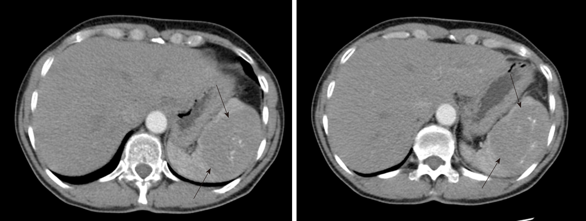
Figure 1 Abdomen computed tomography of a 65-year-old female patient showing a 74 mm × 54 mm solid splenic lesion (marked with arrows).
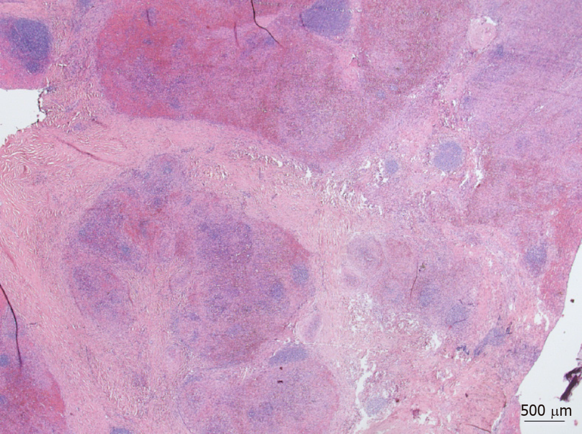
Figure 2 Histological appearance of the first case with typical findings of sclerosing angiomatoid nodular transformation.
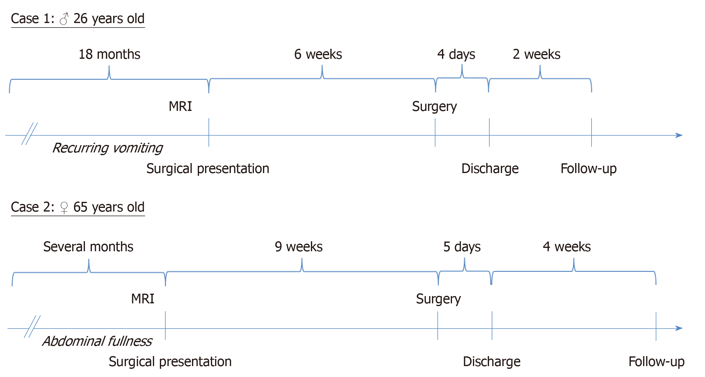
Figure 3 Timeline of both cases.
- Citation: Chikhladze S, Lederer AK, Fichtner-Feigl S, Wittel UA, Werner M, Aumann K. Sclerosing angiomatoid nodular transformation of the spleen, a rare cause for splenectomy: Two case reports. World J Clin Cases 2020; 8(1): 103-109
- URL: https://www.wjgnet.com/2307-8960/full/v8/i1/103.htm
- DOI: https://dx.doi.org/10.12998/wjcc.v8.i1.103


