Copyright
©The Author(s) 2019.
World J Clin Cases. Apr 26, 2019; 7(8): 984-991
Published online Apr 26, 2019. doi: 10.12998/wjcc.v7.i8.984
Published online Apr 26, 2019. doi: 10.12998/wjcc.v7.i8.984
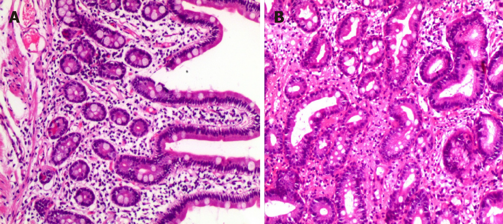
Figure 1 Pathological results of gastric biopsy.
A: Visible mucosal glands are still well differentiated, with many lymphocytes and a small number of eosinophils infiltrating in the stroma observed under a light microscope (magnification, 100×); B: Mild intestinal metaplasia of mucosal glands and a small amount of interstitial lymphocyte, plasma cell, and eosinophil infiltration under a light microscope (magnification, 200×).
Figure 2 Pathological results of colon biopsy.
A: The glandular differentiation is still good and there are many inflammatory cells infiltrating in the interstitial tissues (magnification, 100×); B: Large numbers of lymphocytes and a small amount of eosinophil infiltration (magnification, 200×).
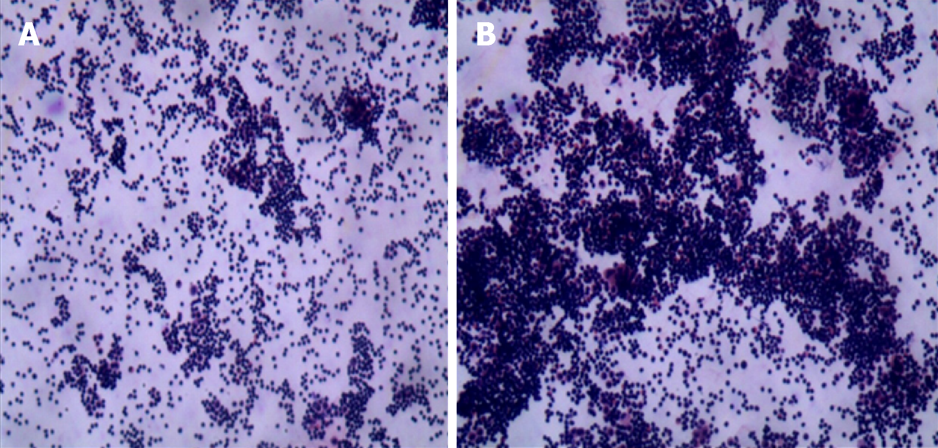
Figure 3 Cytology results of ascites.
A: A large number of lymphocytes can be seen under a light microscope (magnification, 100×); B: A large number of lymphocytes are visible under a light microscope (magnification, 200×). The mesothelial cells, phagocytes, and a few eosinophils can be seen.
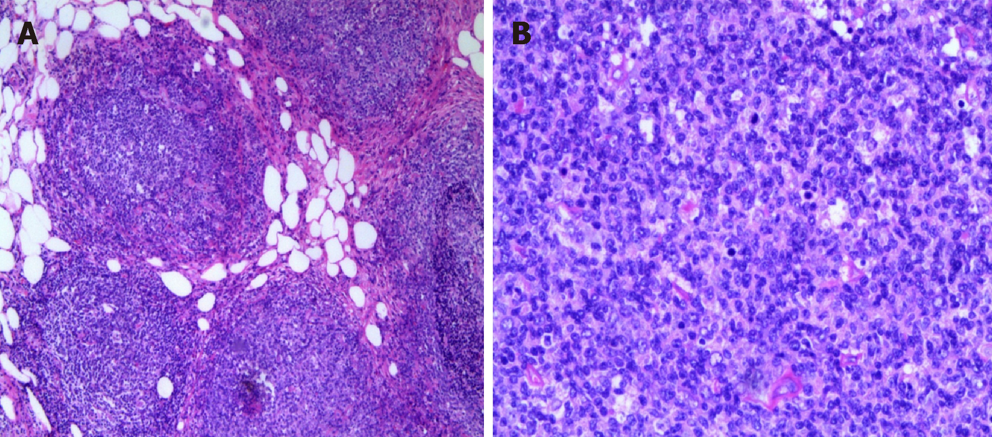
Figure 4 Omental histopathological examination.
A: Lymphoid hyperplasia in omental adipose tissue; B: Some follicles were fused, and no typical cuff was observed. The follicular node was seen under a light microscope (magnification, 200×). The section consists of central cells and centroblasts, and the number of centroblasts is > 15/HPF.
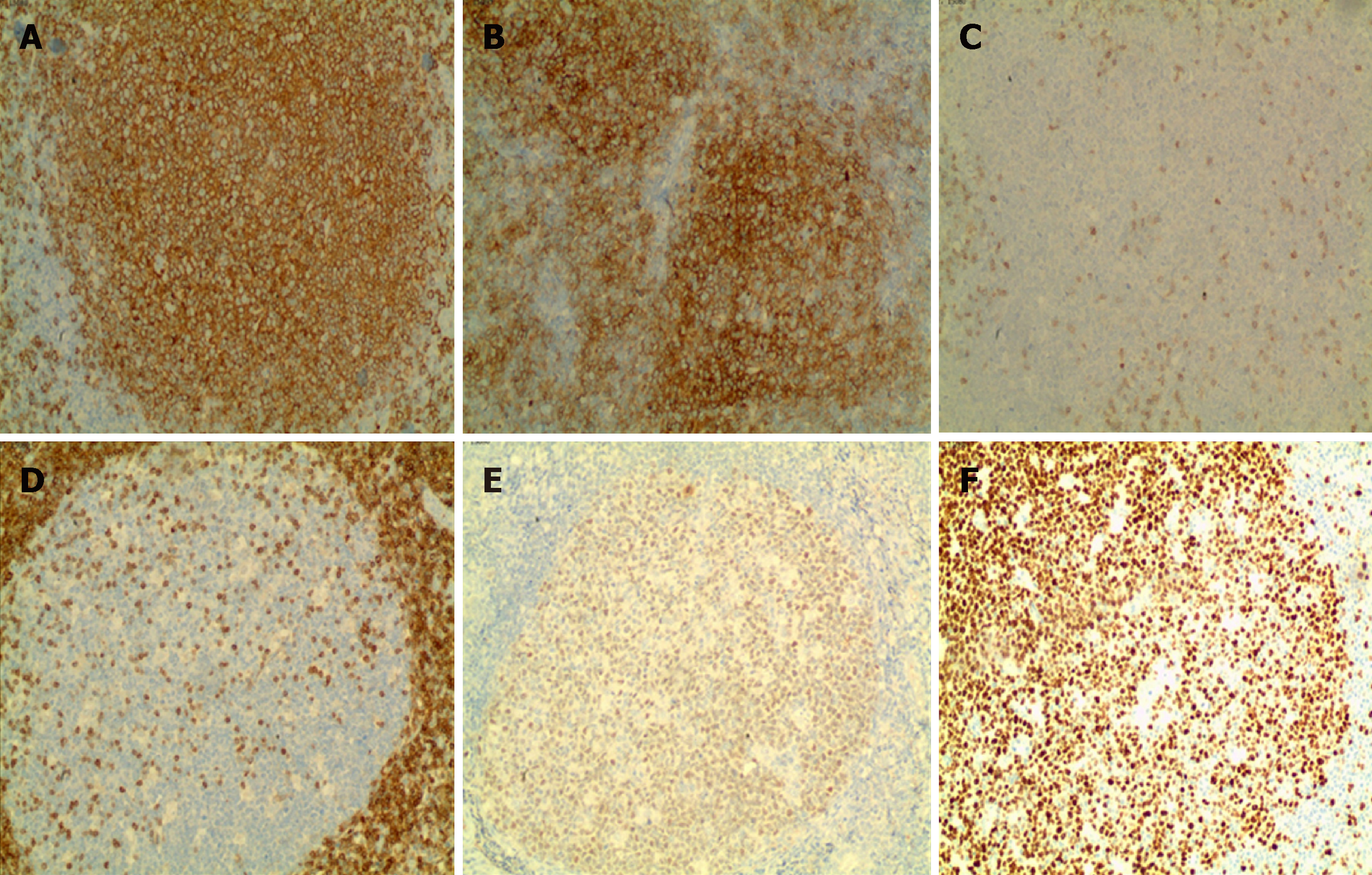
Figure 5 Results of omental immunohistochemistry (100×).
A: Staining for CD20 is diffuse and strongly positive; B: Staining for CD10 is positive; C: Staining for CD3 is negative; D: Staining for Bcl-2 is negative; E: Staining for Bcl-6 is positive; F: Ki-67 positive rate is approximately 60%.
- Citation: Wei C, Xiong F, Yu ZC, Li DF, Luo MH, Liu TT, Li YX, Zhang DG, Xu ZL, Jin HT, Tang Q, Wang LS, Wang JY, Yao J. Diagnosis of follicular lymphoma by laparoscopy: A case report. World J Clin Cases 2019; 7(8): 984-991
- URL: https://www.wjgnet.com/2307-8960/full/v7/i8/984.htm
- DOI: https://dx.doi.org/10.12998/wjcc.v7.i8.984


