Copyright
©The Author(s) 2019.
World J Clin Cases. Mar 26, 2019; 7(6): 798-804
Published online Mar 26, 2019. doi: 10.12998/wjcc.v7.i6.798
Published online Mar 26, 2019. doi: 10.12998/wjcc.v7.i6.798
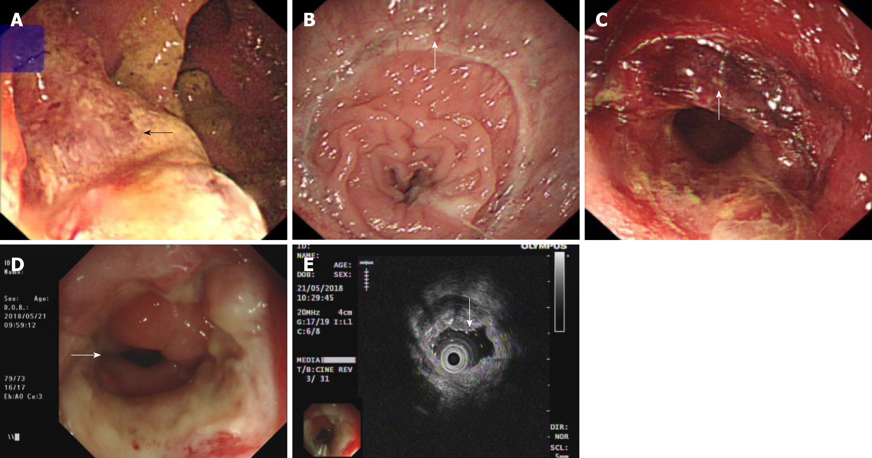
Figure 1 Findings of colonoscopy and endoscopic ultrasonography during treatment.
A: The colonoscopy revealed a 3 cm × 4 cm ulcerated mass in the rectum 4 cm from the anus before surgery; B: After surgery, there was a rectal mucosal inflammation change without residual tumour on colonoscopy; C: The patient underwent photodynamic therapy, a circular piece of mucosal tissue fell off the anus, but the anastomotic stenosis was observed at 5 cm from the anus; D and E: Five years after the operation, there was an intestinal canal stenosis without tumour on endoscopic ultrasonography.
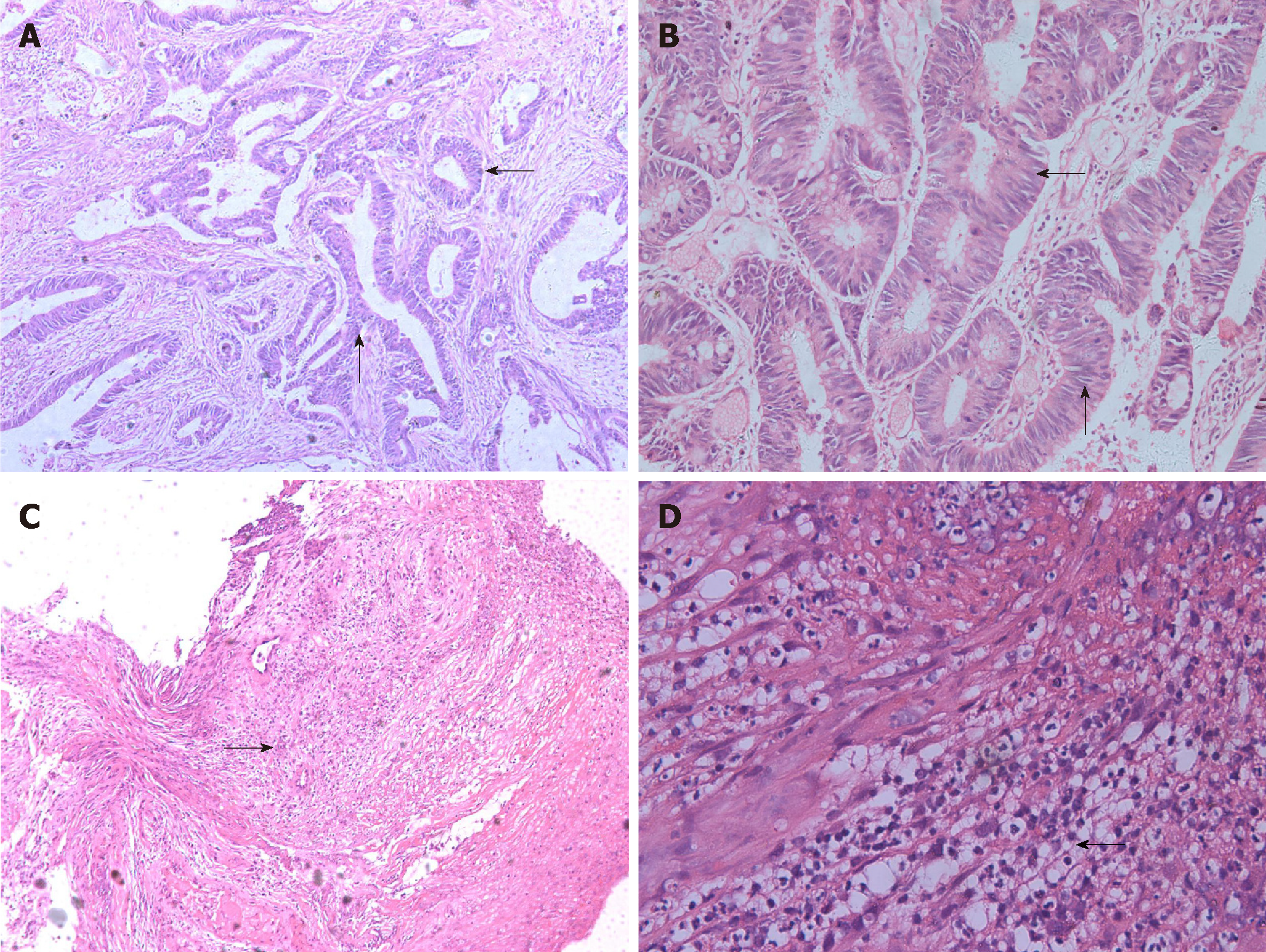
Figure 2 Pathological changes in the patient with positive microscopic distal margins after photodynamic therapy.
A and B: Postoperative histopathologic examination revealed that the tumour was a moderately differentiated rectal adenocarcinoma and a partial mucinous adenocarcinoma; the TNM stage was T2N0M0, with no cancer in the incision margin and no metastasis (0/4) to the lymph nodes. Unfortunately, cancer was detected on the lateral edge of the operated area; C and D: Five years after the operation, microscopic examination of a biopsy at the rectal anastomosis showed the formation of inflammatory necrotic lesions and the formation of ulcerative lesions in the inflammatory granulation tissue. No malignancy was found in multiple sections.
- Citation: Zhang SQ, Liu KJ, Yao HL, Lei SL, Lei ZD, Yi WJ, Xiong L, Zhao H. Photodynamic therapy as salvage therapy for residual microscopic cancer after ultra-low anterior resection: A case report. World J Clin Cases 2019; 7(6): 798-804
- URL: https://www.wjgnet.com/2307-8960/full/v7/i6/798.htm
- DOI: https://dx.doi.org/10.12998/wjcc.v7.i6.798


