Copyright
©The Author(s) 2019.
World J Clin Cases. Mar 26, 2019; 7(6): 765-772
Published online Mar 26, 2019. doi: 10.12998/wjcc.v7.i6.765
Published online Mar 26, 2019. doi: 10.12998/wjcc.v7.i6.765
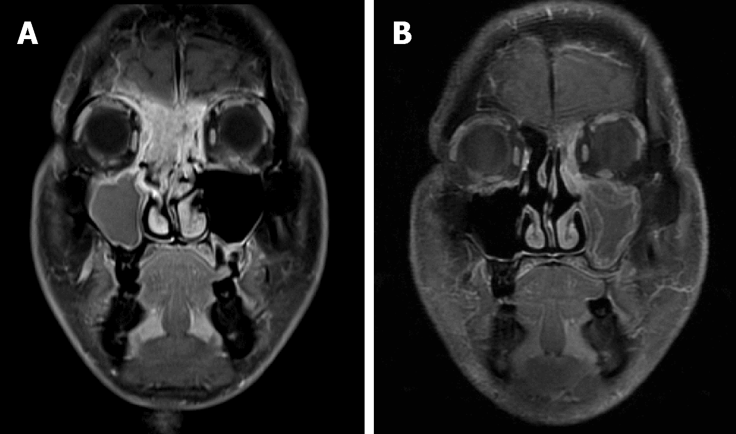
Figure 1 Imaging features in case 1.
A: Magnetic resonance imaging (MRI) of the head on admission. The tumor had widely invaded the paranasal sinuses, the right orbit and the cranial base; B: MRI after concurrent chemoradiotherapy showed a complete response of the tumor.
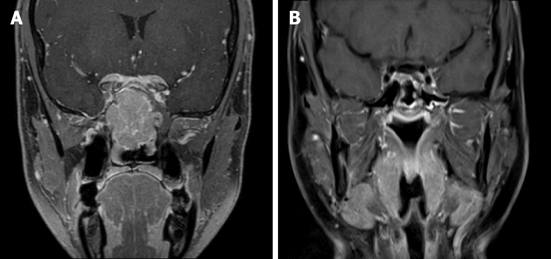
Figure 2 Imaging features in case 2.
A: On admission, the tumor involved the paranasal sinuses and the cranial base; B: After the treatment, a complete response of the disease was observed by magnetic resonance imaging.
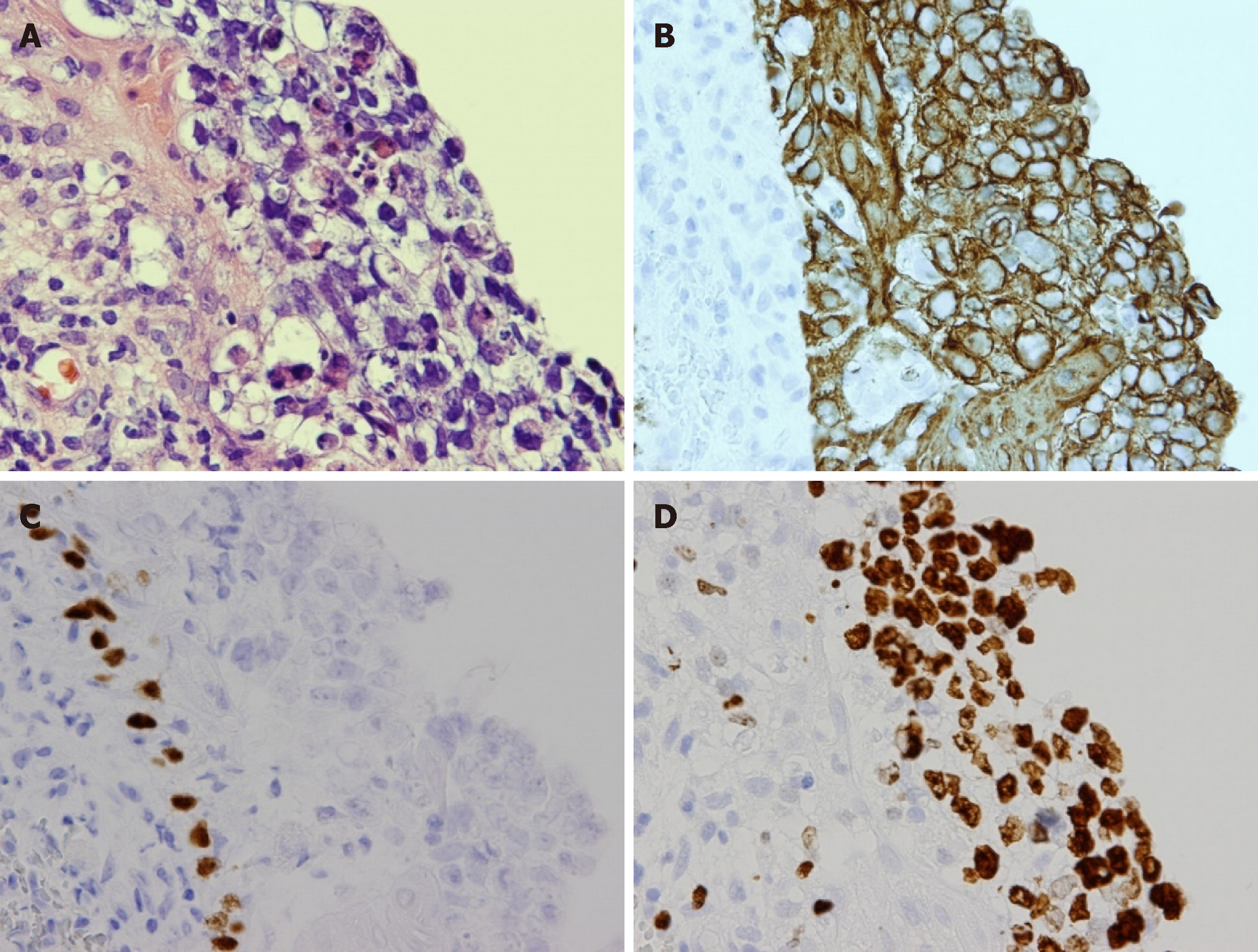
Figure 3 Pathological findings in case 1.
A: Pleomorphic tumor cells with hyperchromatic nuclei, clear cytoplasm and poorly-defined cell borders were seen (hematoxylin and eosin, Ã 400); B: Immunohistochemically, the tumor cells were positive for AE1/3; C: The tumor cells were negative for p40; D: A high expression of Ki67 was also noted.
- Citation: Watanabe S, Honma Y, Murakami N, Igaki H, Mori T, Hirano H, Okita N, Shoji H, Iwasa S, Takashima A, Kato K, Kobayashi K, Matsumoto F, Yoshimoto S, Itami J, Boku N. Induction chemotherapy with docetaxel, cisplatin and fluorouracil followed by concurrent chemoradiotherapy for unresectable sinonasal undifferentiated carcinoma: Two cases of report. World J Clin Cases 2019; 7(6): 765-772
- URL: https://www.wjgnet.com/2307-8960/full/v7/i6/765.htm
- DOI: https://dx.doi.org/10.12998/wjcc.v7.i6.765


