Copyright
©The Author(s) 2019.
World J Clin Cases. Mar 26, 2019; 7(6): 759-764
Published online Mar 26, 2019. doi: 10.12998/wjcc.v7.i6.759
Published online Mar 26, 2019. doi: 10.12998/wjcc.v7.i6.759
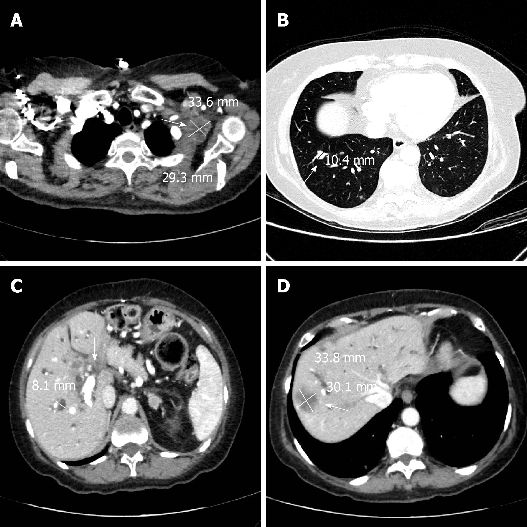
Figure 1 A total body computed tomography scan confirmed the metastases in the lungs, chest, abdominal lymph nodes, liver and hepatic hilar area.
A: Arrow showing pathological lymphadenopathy in the left axilla (34 mm × 29 mm); B: Arrow showing secondary lesion of the pulmonary parenchyma in the lower right lobe (10 mm); C: Arrow showing pathological tissue enveloping the hepatic hilum; dilated intrahepatic bile ducts in right lobe (8 mm); D: Arrow showing secondary lesion to the VIII segment of the liver.
- Citation: Monti M, Torri A, Amadori E, Rossi A, Bartolini G, Casadei C, Frassineti GL. Aeromonas veronii biovar veronii and sepsis-infrequent complication of biliary drainage placement: A case report. World J Clin Cases 2019; 7(6): 759-764
- URL: https://www.wjgnet.com/2307-8960/full/v7/i6/759.htm
- DOI: https://dx.doi.org/10.12998/wjcc.v7.i6.759


