Copyright
©The Author(s) 2019.
World J Clin Cases. Mar 26, 2019; 7(6): 727-733
Published online Mar 26, 2019. doi: 10.12998/wjcc.v7.i6.727
Published online Mar 26, 2019. doi: 10.12998/wjcc.v7.i6.727
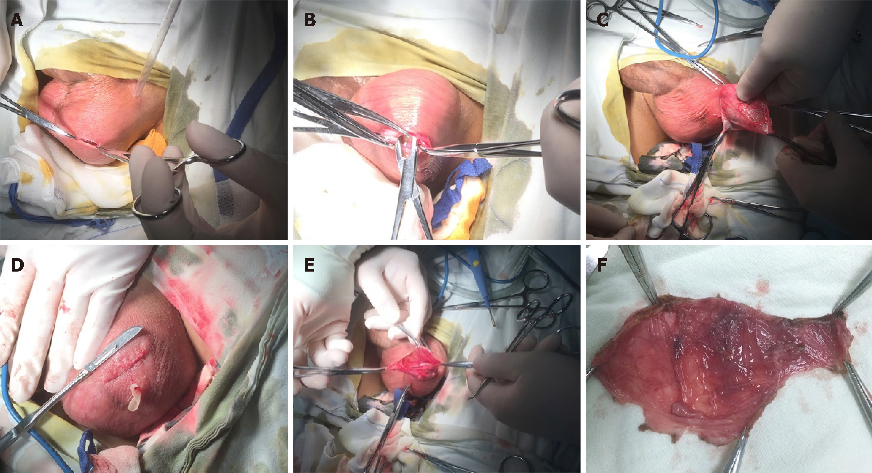
Figure 1 Surgical protocol.
A: A 2-cm incision is made in the transverse and anterior skin of the scrotum; B: The sheath cavity is exposed; C: The forefinger of the left hand is used to extend into the sheath cavity to assist the separation and protect the contents of the scrotum from damage; D: The sheath tissue is separated as much as possible until the intended target size is reached; E: The small incision is closed with absorbable sutures; F: The resected sheath tissue is removed and routinely sent for pathological examination.
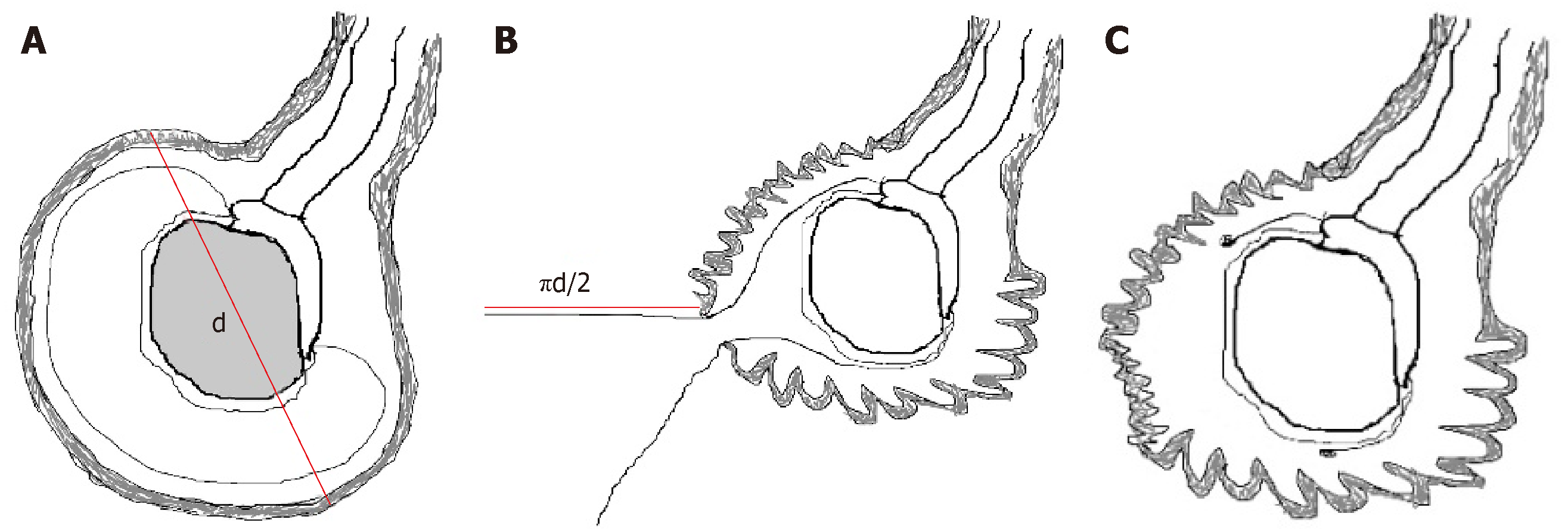
Figure 2 Diagram of the surgical protocol.
A: The maximum diameter of the effusion (d) is measured by preoperative ultrasound; B: The maximum diameter of the sheath that would be peeled off through the small incision is approximately πd/2; C: A sufficient portion of the sheath is removed to prevent recurrence of hydrocele.
- Citation: Lin L, Hong HS, Gao YL, Yang JR, Li T, Zhu QG, Ye LF, Wei YB. Individualized minimally invasive treatment for adult testicular hydrocele: A pilot study. World J Clin Cases 2019; 7(6): 727-733
- URL: https://www.wjgnet.com/2307-8960/full/v7/i6/727.htm
- DOI: https://dx.doi.org/10.12998/wjcc.v7.i6.727


