Copyright
©The Author(s) 2019.
World J Clin Cases. Feb 26, 2019; 7(4): 516-524
Published online Feb 26, 2019. doi: 10.12998/wjcc.v7.i4.516
Published online Feb 26, 2019. doi: 10.12998/wjcc.v7.i4.516
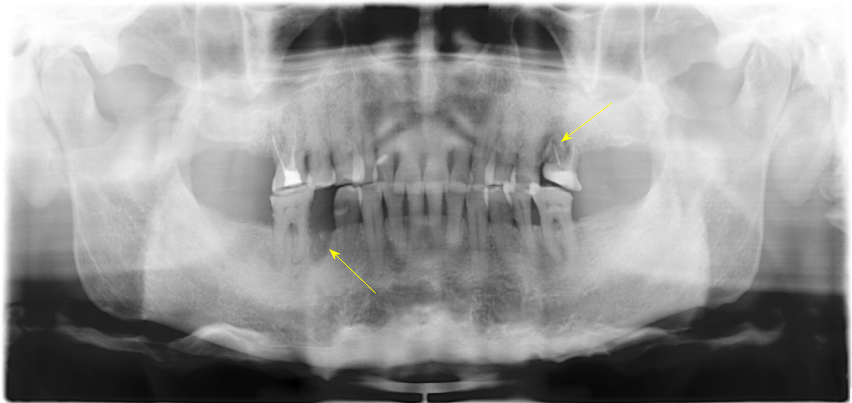
Figure 1 Panoramic radiograph indicating a fresh extraction socket in #45 and suspected root fracture in #26.
Multiple molar teeth (#45, #17, #27, #37, and #47) were lost.
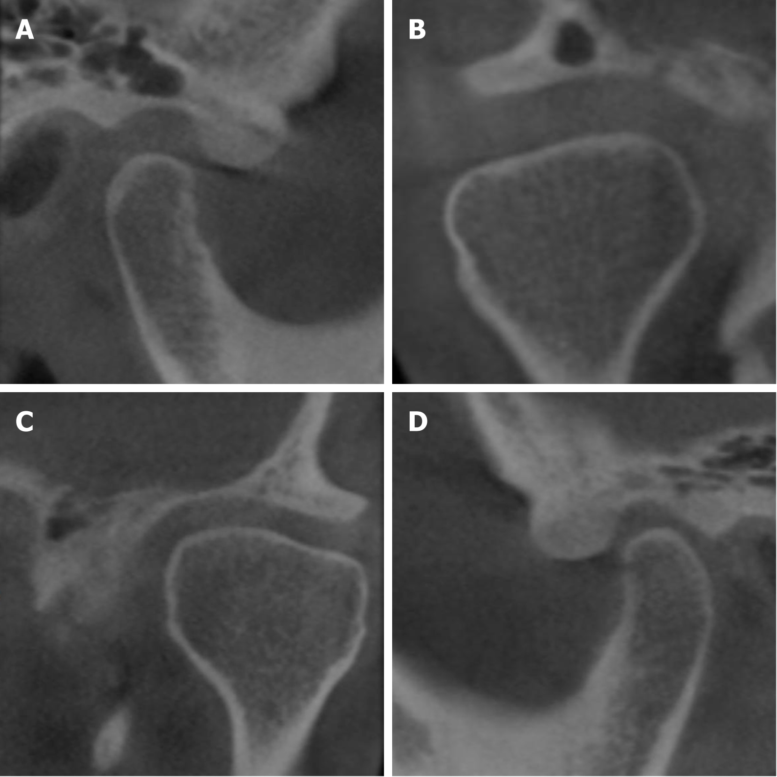
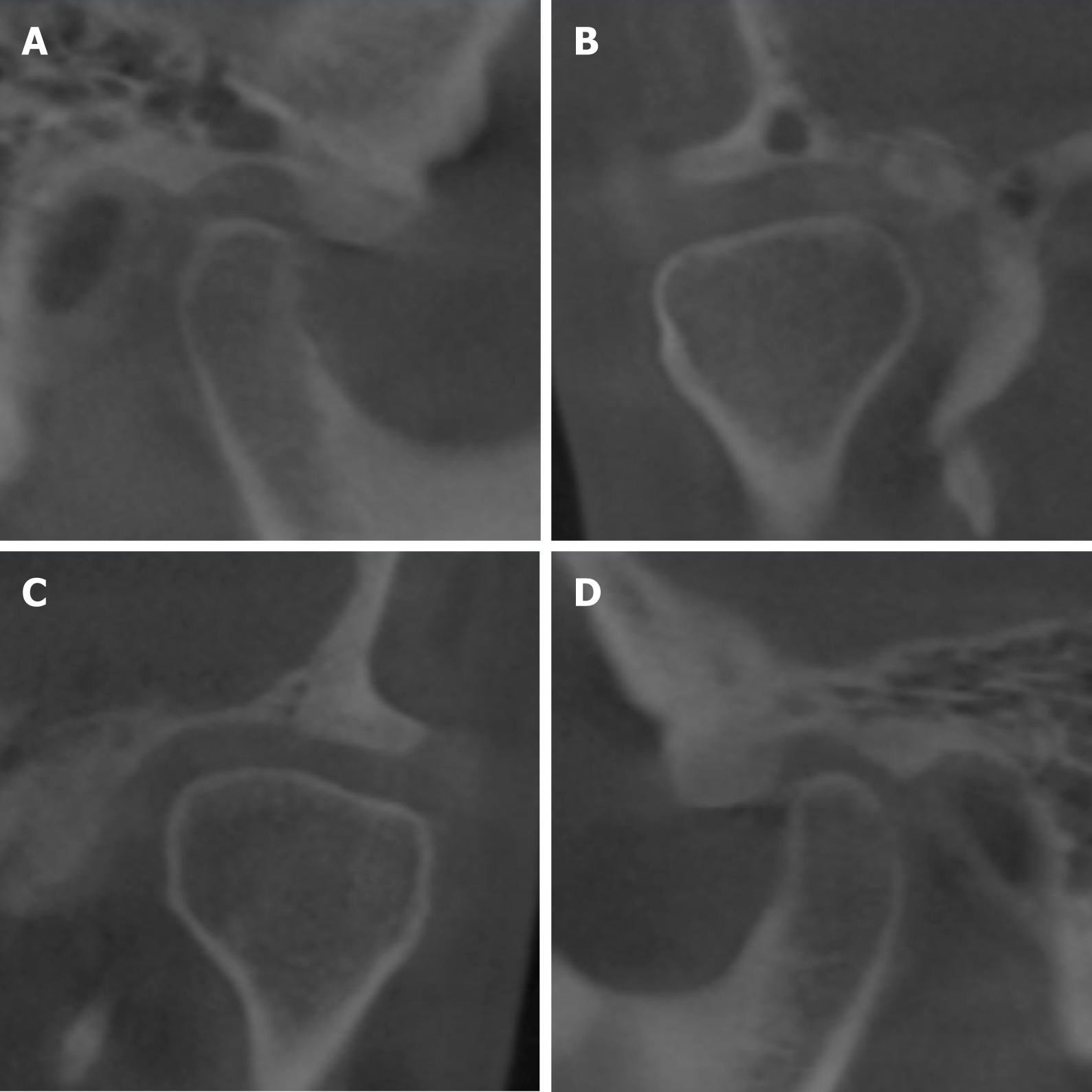
Figure 2 Cone-beam computed tomography examination of the temporomandibular joint.
A: Sagittal projection of the right temporomandibular joint (TMJ) showing increased posterior joint space; B: Coronal projection of the right TMJ showing increased medial and lateral joint space; C: Coronal projection of the left TMJ; D: Sagittal projection of the left TMJ.
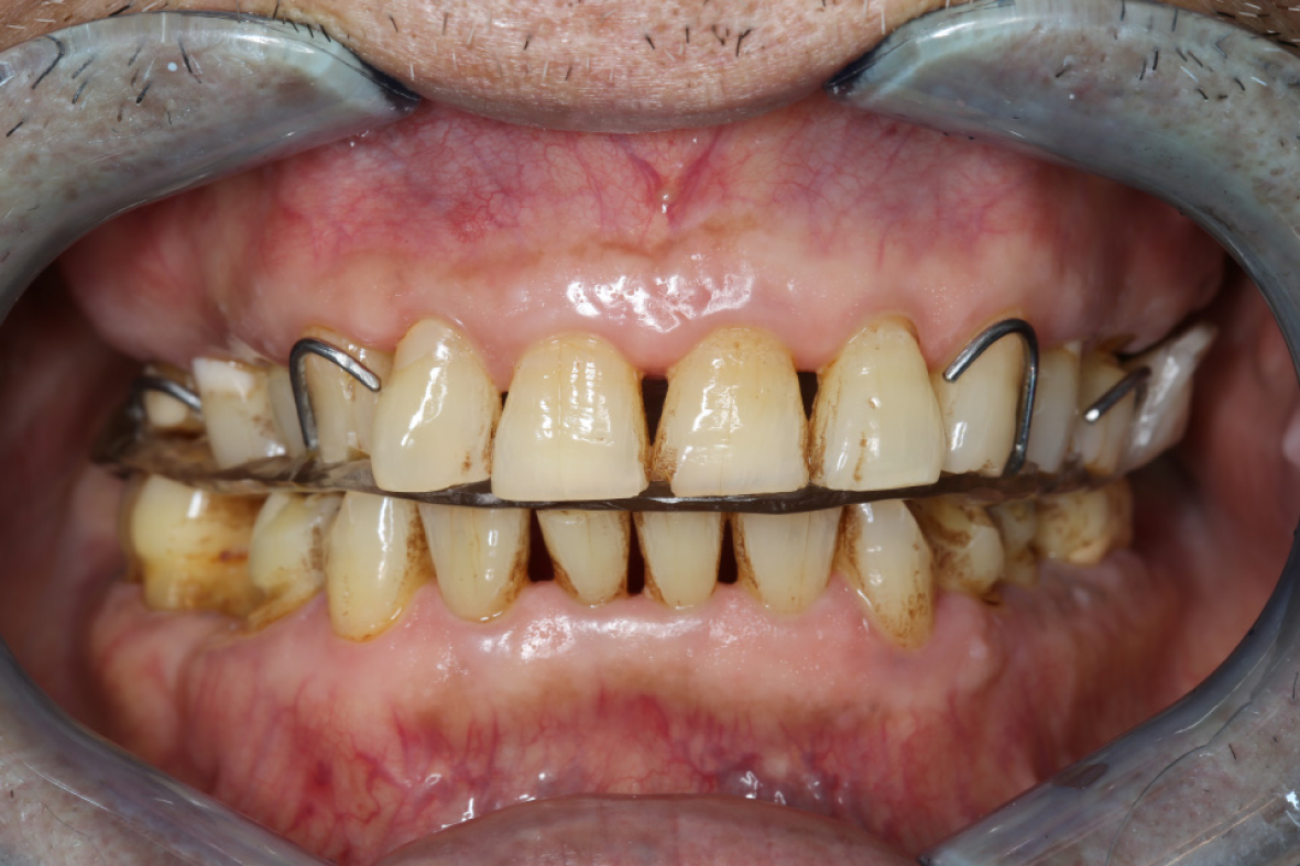
Figure 3 Maxillary buccal-pterygoid splint with reinforced wire fused into the bilateral buccal-pterygoid pads was positioned in the mouth.
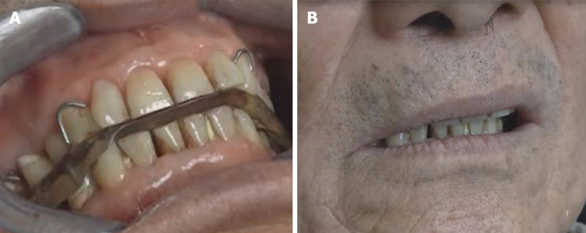
Figure 4 Video recordings demonstrating stable control of tooth grinding.
A: Wearing the splint, his tooth grinding activity was limited to a relatively smaller range; B: Removing the splint, involuntary tooth grinding gradually re-started.
Figure 5 Bilateral buccal-pterygoid pads were removed after daytime tooth grinding symptoms were almost completely controlled.
Figure 6 Follow-up video recordings demonstrating almost completely ceased tooth grinding and clenching.
A: Wearing the splint, his tooth grinding completely ceased with very mild symptoms of clenching; B: Removing the splint, his tooth grinding did not recur. Slight relapse of clenching can be controlled consciously.
Figure 7 Seven-year follow-up cone-beam computed tomography images indicating no change when compared with the previous images.
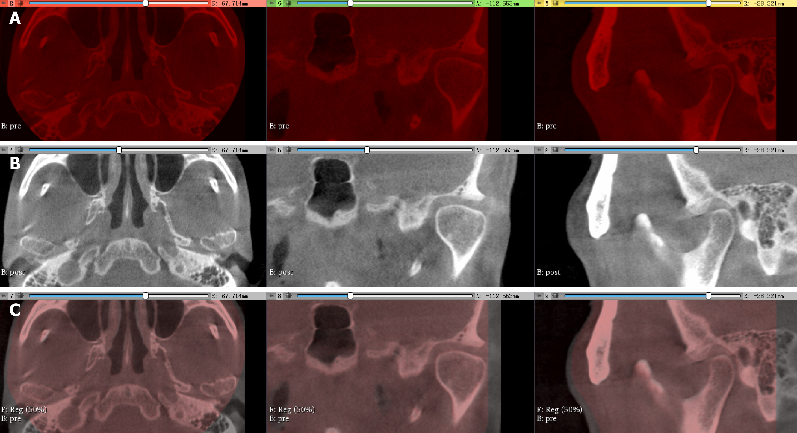
Figure 8 Registration of the pre- and post-cone-beam computed tomography images.
A: Cone-beam computed tomography (CBCT) images pre-treatment are labeled red; B: Seven-year follow-up CBCT images post-treatment are labeled gray; C: General registration was performed to superimpose the pre-CBCT and post-CBCT images. Pre-CBCT images served as fixed images with a 50% transparency while post-CBCT images as moving images. A perfect match of the whole images revealed no significant longitudinal changes of the temporomandibular joint.
- Citation: Chen JM, Yan Y. Long-term follow-up of a patient with venlafaxine-induced diurnal bruxism treated with an occlusal splint: A case report. World J Clin Cases 2019; 7(4): 516-524
- URL: https://www.wjgnet.com/2307-8960/full/v7/i4/516.htm
- DOI: https://dx.doi.org/10.12998/wjcc.v7.i4.516


