Copyright
©The Author(s) 2019.
World J Clin Cases. Dec 26, 2019; 7(24): 4377-4383
Published online Dec 26, 2019. doi: 10.12998/wjcc.v7.i24.4377
Published online Dec 26, 2019. doi: 10.12998/wjcc.v7.i24.4377
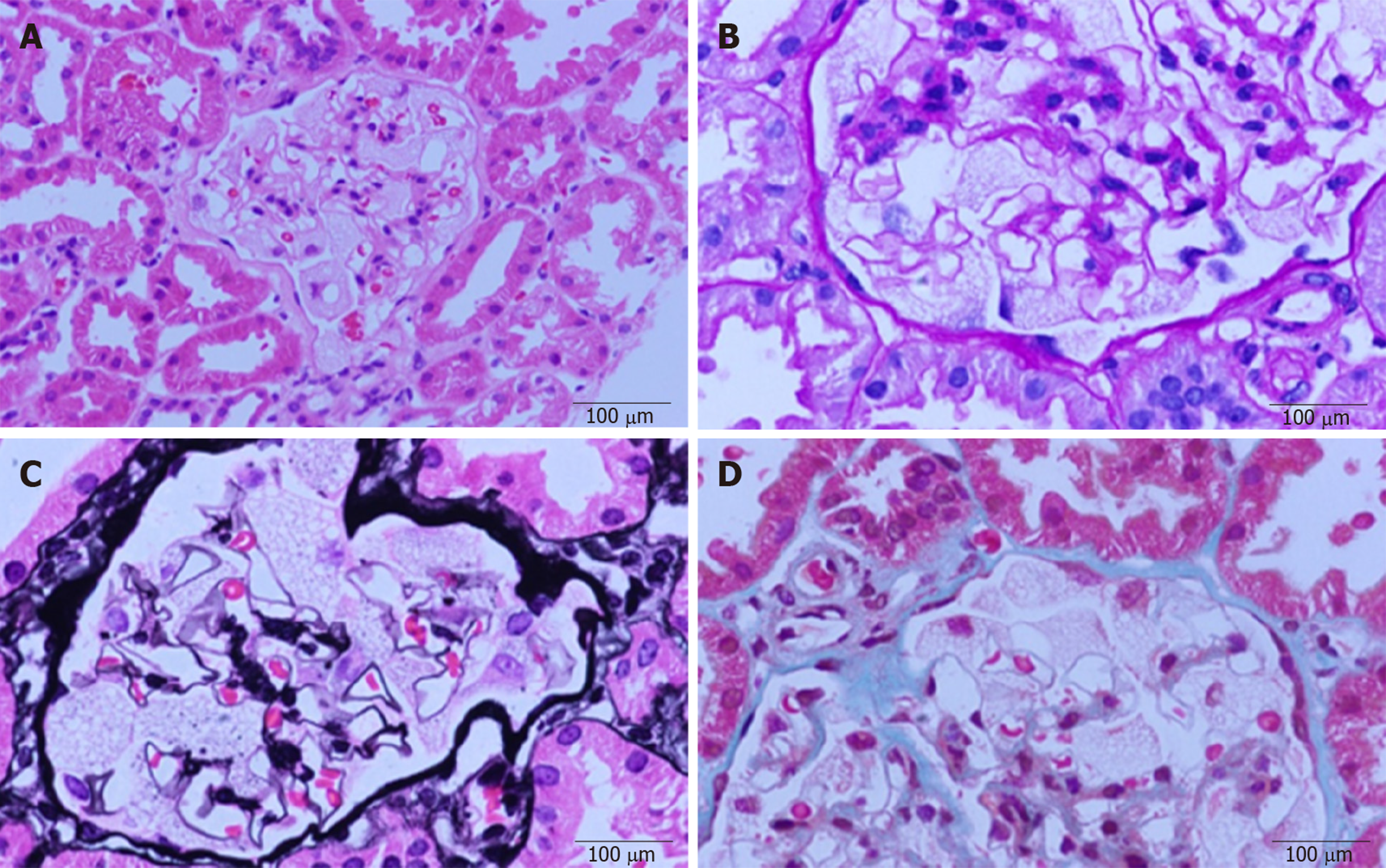
Figure 1 Light microscopic images.
Diffuse enlargement and vacuolar degeneration of glomerular visceral epithelial cells are seen. A: Hematoxylin-eosin staining; B: Periodic acid-Schiff staining; C: Periodic acid-silver methenamine staining; and D: Masson staining.
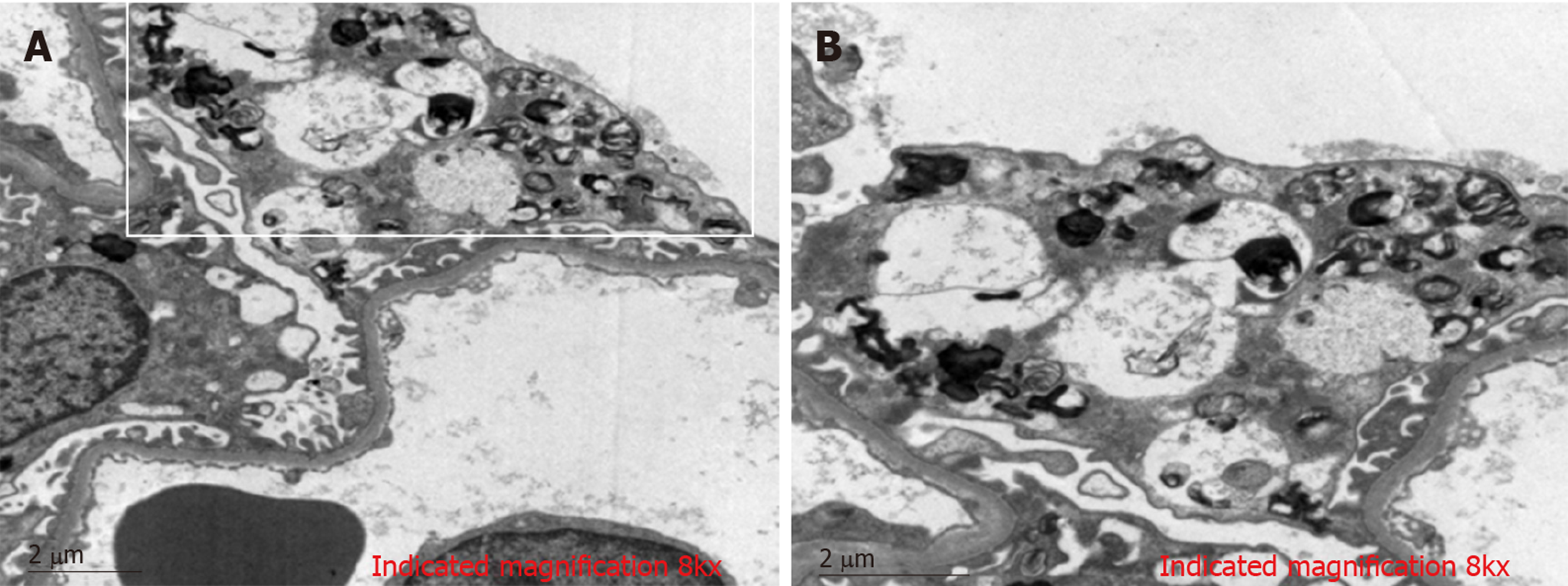
Figure 2 Electron microscopic images.
Vacuoles with dense lamellated structures are seen in glomerular visceral epithelial cells. Such structures are called zebra and myeloid bodies. Podocyte foot processes appear to be effaced (image B is an enlargement of the part of image A within the white box). Image magnifications are specified at the bottom of each micrograph.
- Citation: Wu SZ, Liang X, Geng J, Zhang MB, Xie N, Su XY. Hydroxychloroquine-induced renal phospholipidosis resembling Fabry disease in undifferentiated connective tissue disease: A case report. World J Clin Cases 2019; 7(24): 4377-4383
- URL: https://www.wjgnet.com/2307-8960/full/v7/i24/4377.htm
- DOI: https://dx.doi.org/10.12998/wjcc.v7.i24.4377


