Copyright
©The Author(s) 2019.
World J Clin Cases. Dec 26, 2019; 7(24): 4366-4376
Published online Dec 26, 2019. doi: 10.12998/wjcc.v7.i24.4366
Published online Dec 26, 2019. doi: 10.12998/wjcc.v7.i24.4366
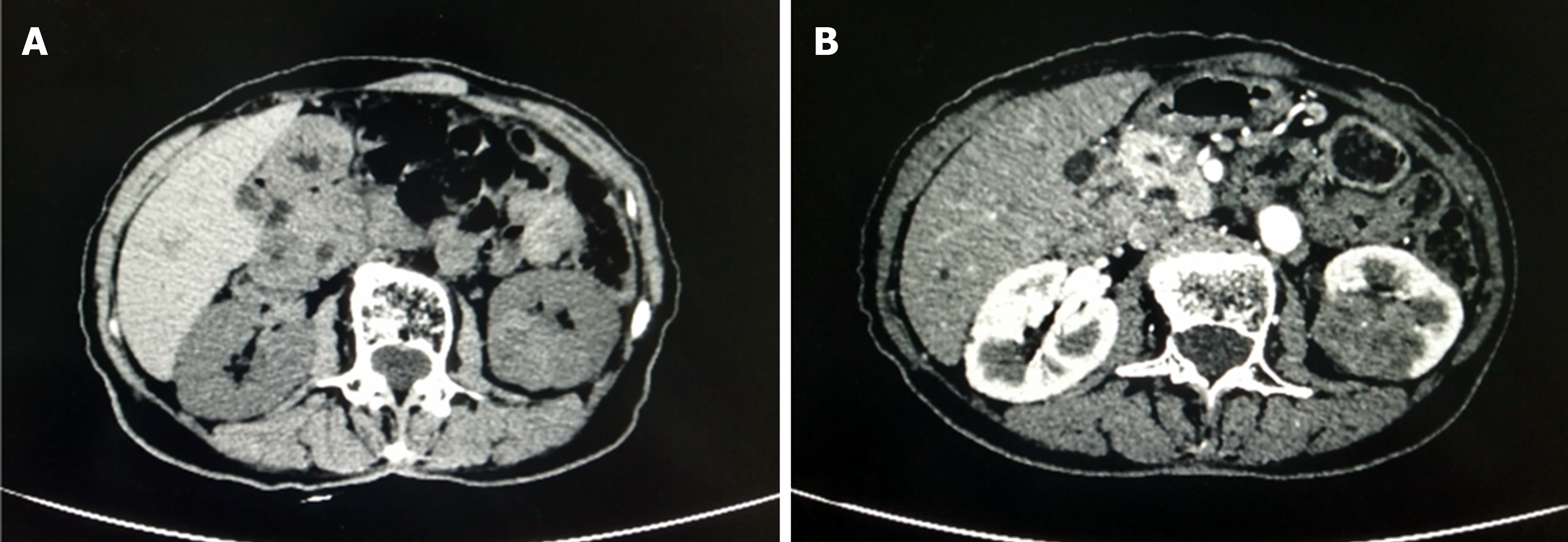
Figure 1 Pre-operative computed tomography images.
A: Computed tomography (CT) image showing a 4.1 cm × 3.2 cm mass in the middle pole of the left kidney. B: Contrast-enhanced CT image showing slight enhancement.
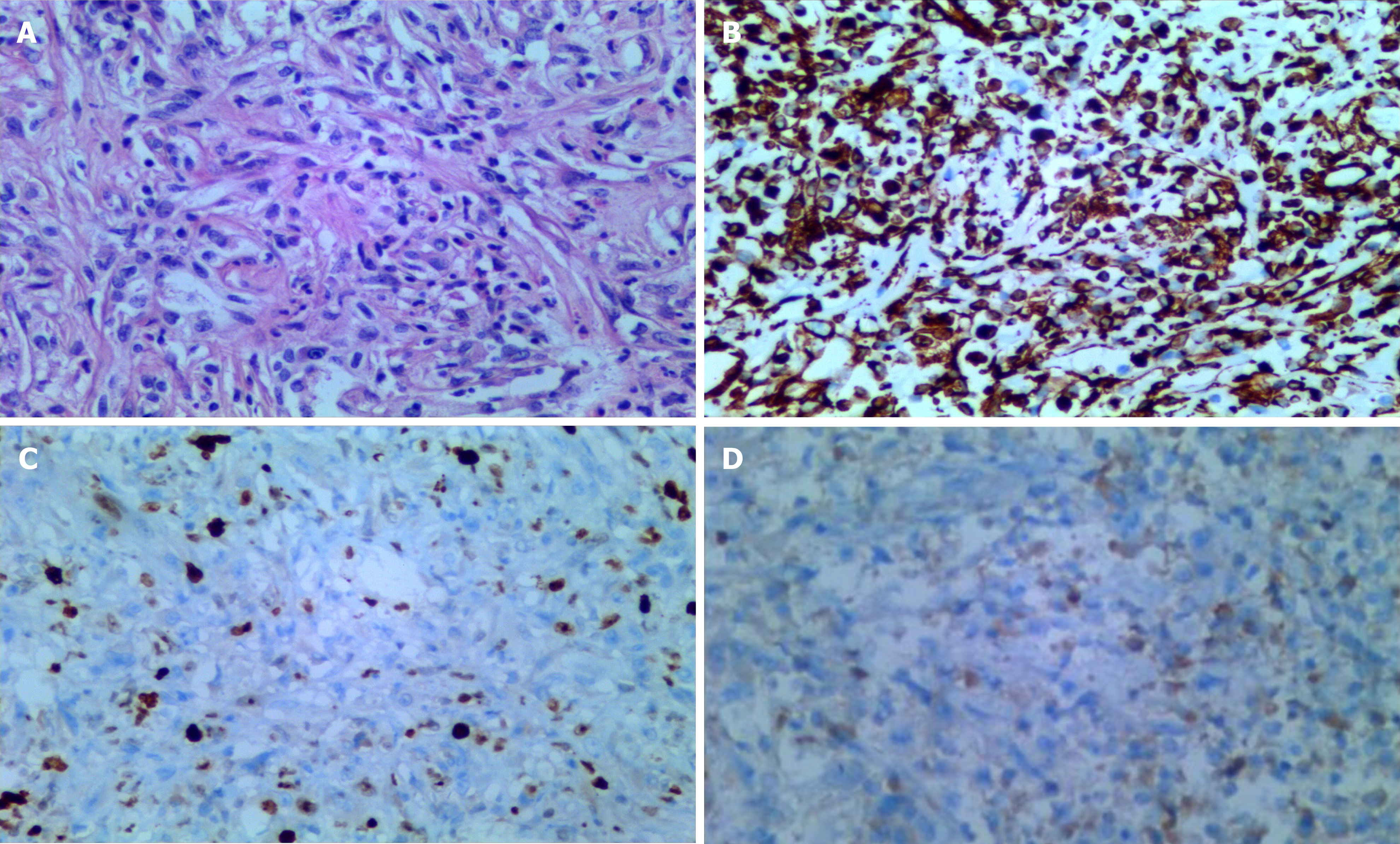
Figure 2 Microscopic findings and immunohistochemical analysis results.
A: Photomicrograph showing abundant spindle cells and collagen with infiltrating lymphocytes and plasma cells (hematoxylin-eosin staining; magnification, ×200); B-D: Immunohistochemically, the tumor cells were positive for (B) vimentin, (C) Ki-67, and (D) CK (magnification, ×200).
- Citation: Zhang GH, Guo XY, Liang GZ, Wang Q. Kidney inflammatory myofibroblastic tumor masquerading as metastatic malignancy: A case report and literature review. World J Clin Cases 2019; 7(24): 4366-4376
- URL: https://www.wjgnet.com/2307-8960/full/v7/i24/4366.htm
- DOI: https://dx.doi.org/10.12998/wjcc.v7.i24.4366


