Copyright
©The Author(s) 2019.
World J Clin Cases. Dec 26, 2019; 7(24): 4299-4306
Published online Dec 26, 2019. doi: 10.12998/wjcc.v7.i24.4299
Published online Dec 26, 2019. doi: 10.12998/wjcc.v7.i24.4299
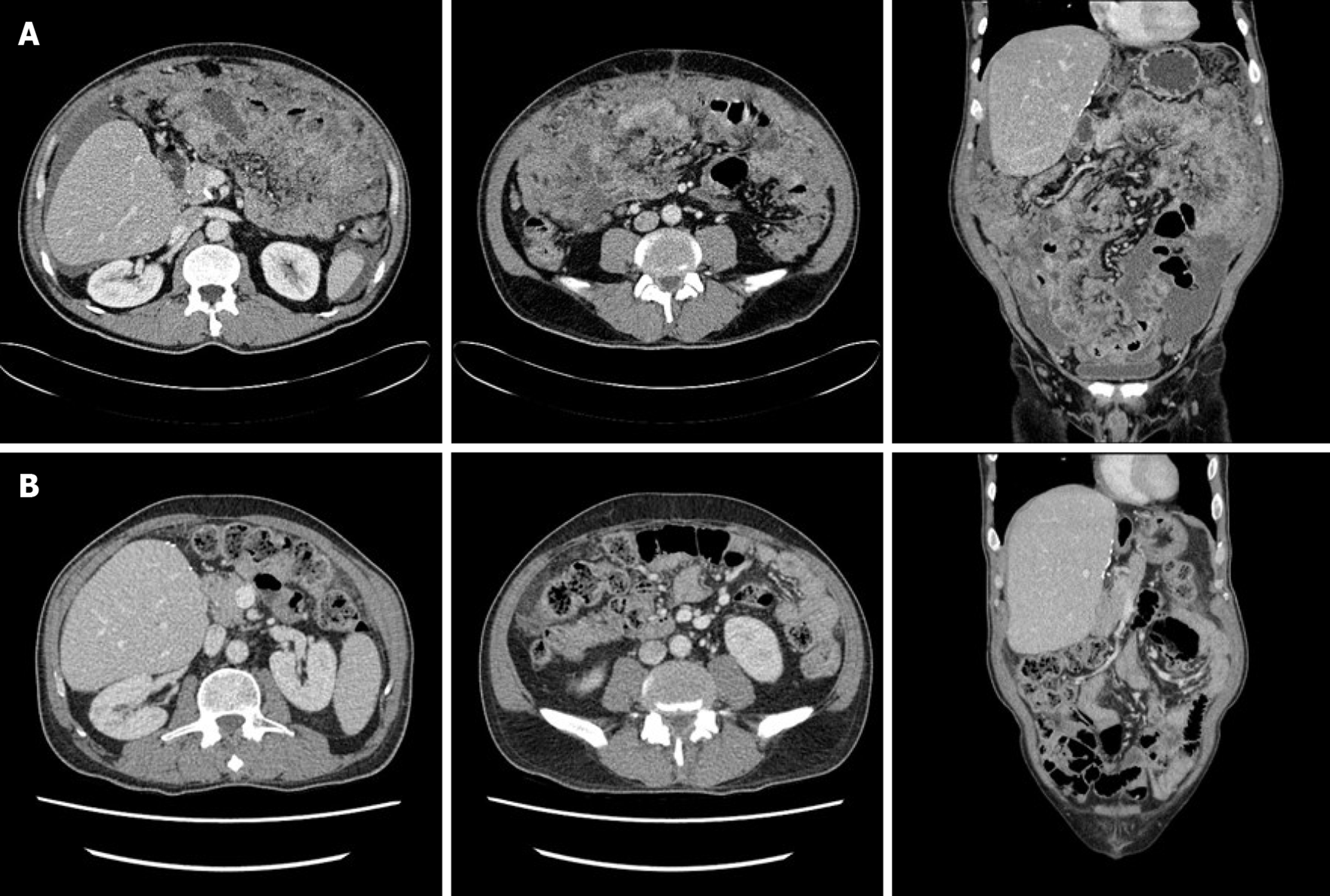
Figure 1 Abdominopelvic computed tomography.
A: Computed tomography (CT) depicting a large volume of ascites and diffuse peritoneal infiltrative lesions at the mesentery and omentum, but no mass-like lesions in the gastrointestinal tract and no bowel obstruction; B: Post-chemotherapy CT depicting no signs of omental mass or ascites, but mild haziness in the omental fat.
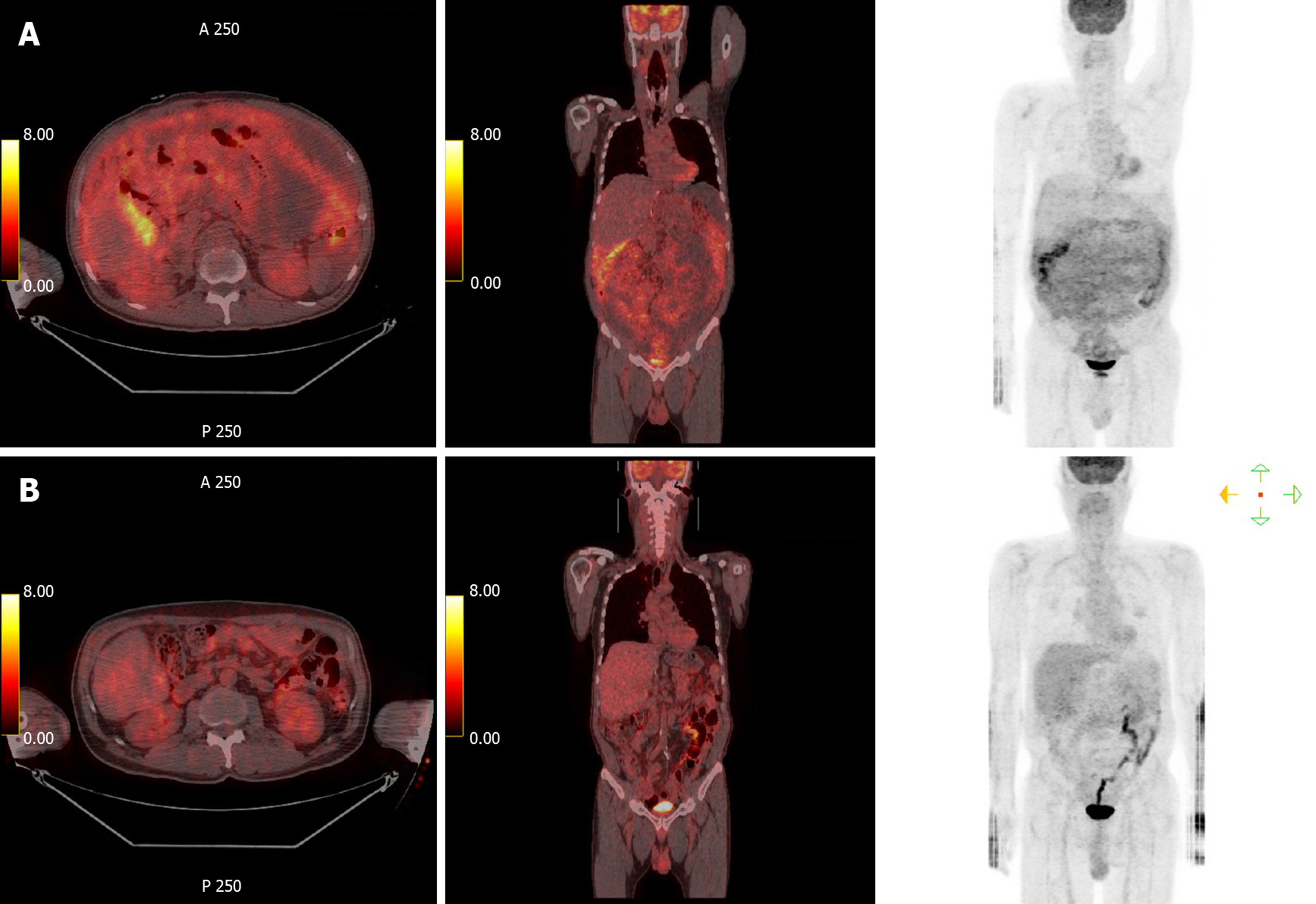
Figure 2 Positron emission tomography-computed tomography.
A: Positron emission tomography (PET)-computed tomography (CT) did not depict abnormal hypermetabolism in solid organs, lymph nodes, or digestive organs, but it did depict diffuse hypermetabolism in the peritoneum and omentum; B: Post-chemotherapy PET-CT depicted loss of diffuse hypermetabolic lesion in the peritoneum.
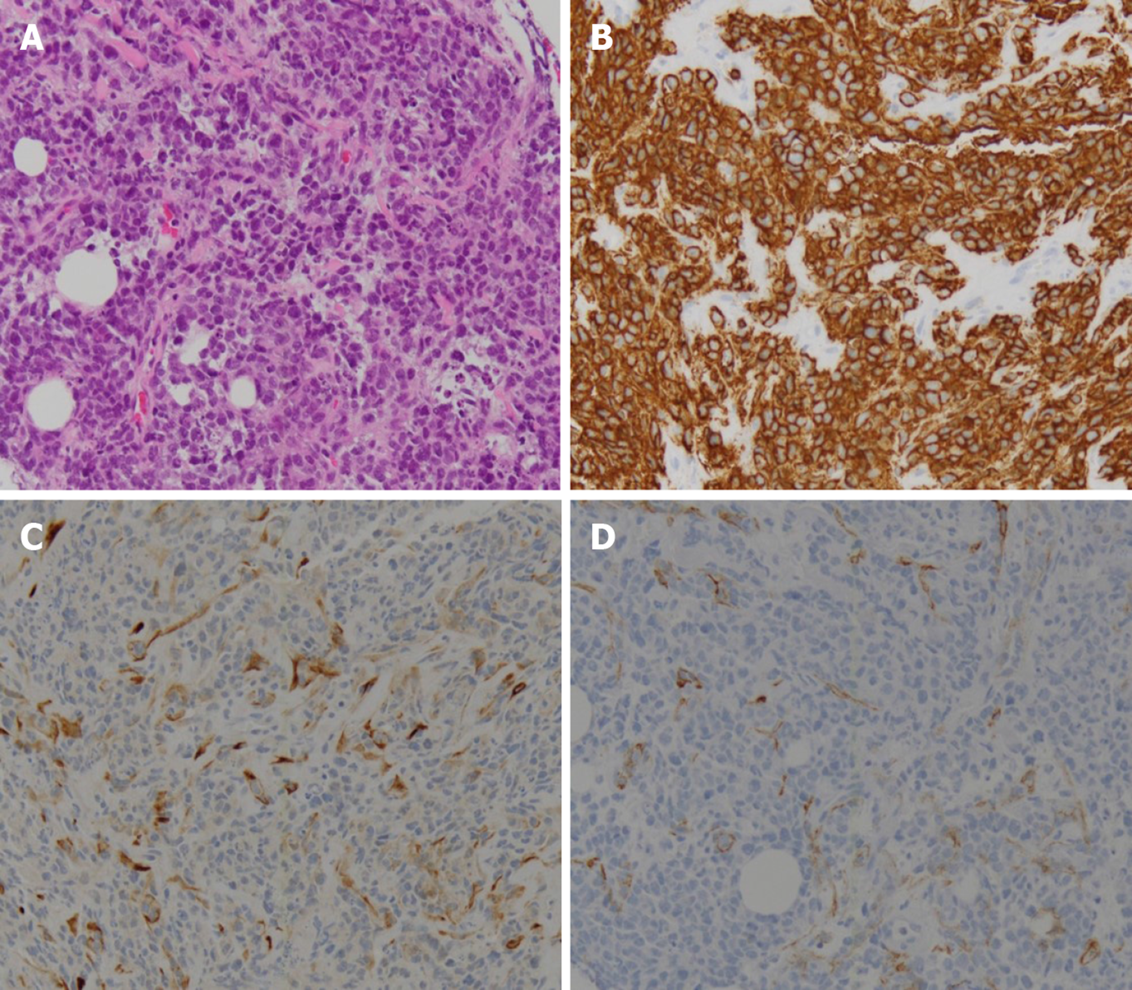
Figure 3 Pathologic findings.
Highly pleomorphic, atypical, small, round, blue cells were observed infiltrating the fibrocollagenous tissue (A). On immunohistochemical testing, these cells were positive for CD20 (B) and LCA, but they were negative for the mesothelial cell marker D2-40 (C) and the epithelial cell marker cytokeratin (D).
- Citation: Kim HB, Hong R, Na YS, Choi WY, Park SG, Lee HJ. Isolated peritoneal lymphomatosis defined as post-transplant lymphoproliferative disorder after a liver transplant: A case report. World J Clin Cases 2019; 7(24): 4299-4306
- URL: https://www.wjgnet.com/2307-8960/full/v7/i24/4299.htm
- DOI: https://dx.doi.org/10.12998/wjcc.v7.i24.4299


