Copyright
©The Author(s) 2019.
World J Clin Cases. Dec 26, 2019; 7(24): 4270-4276
Published online Dec 26, 2019. doi: 10.12998/wjcc.v7.i24.4270
Published online Dec 26, 2019. doi: 10.12998/wjcc.v7.i24.4270
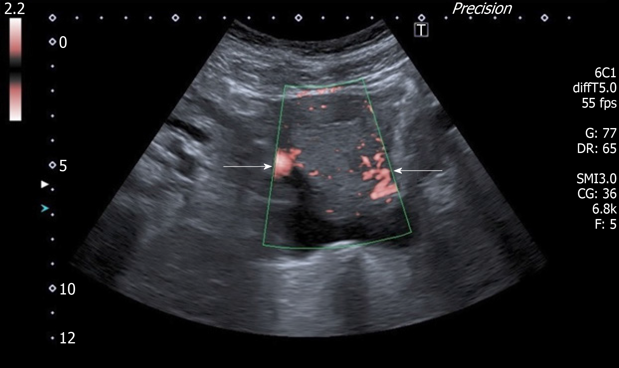
Figure 1 Doppler ultrasound scan of the uterine graft showing perfusion present on both sides of the graft.
Uterine arteries (white arrows).
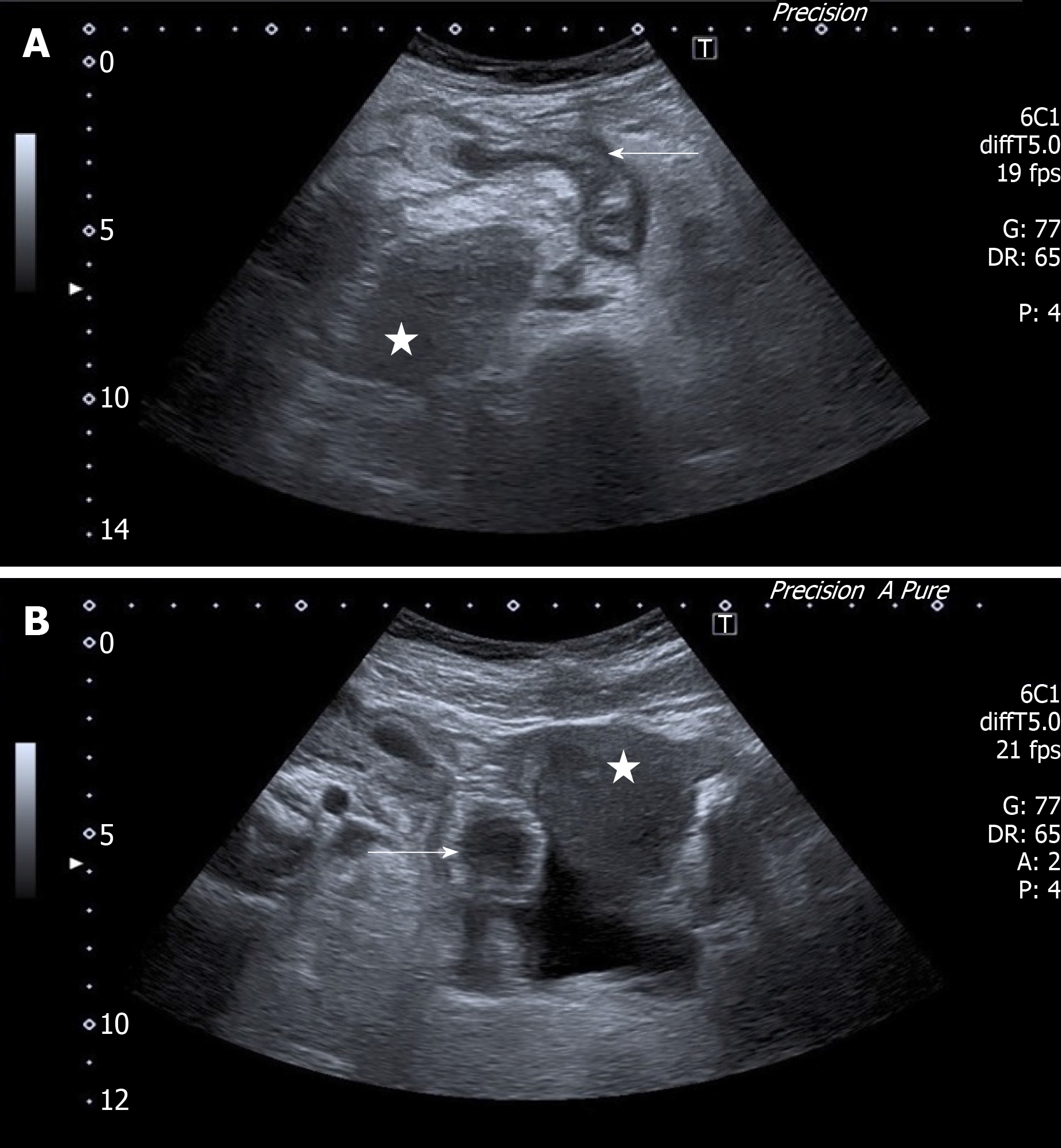
Figure 2 Ultrasound scan.
A: A suspected inflamed appendix next to the uterine graft. Suspected inflamed appendix in a longitudinal view (arrow); the uterine graft (star); B: A suspected inflamed appendix next to the uterine graft. Suspected appendicitis in a transversal view (arrow); the uterine graft (star).
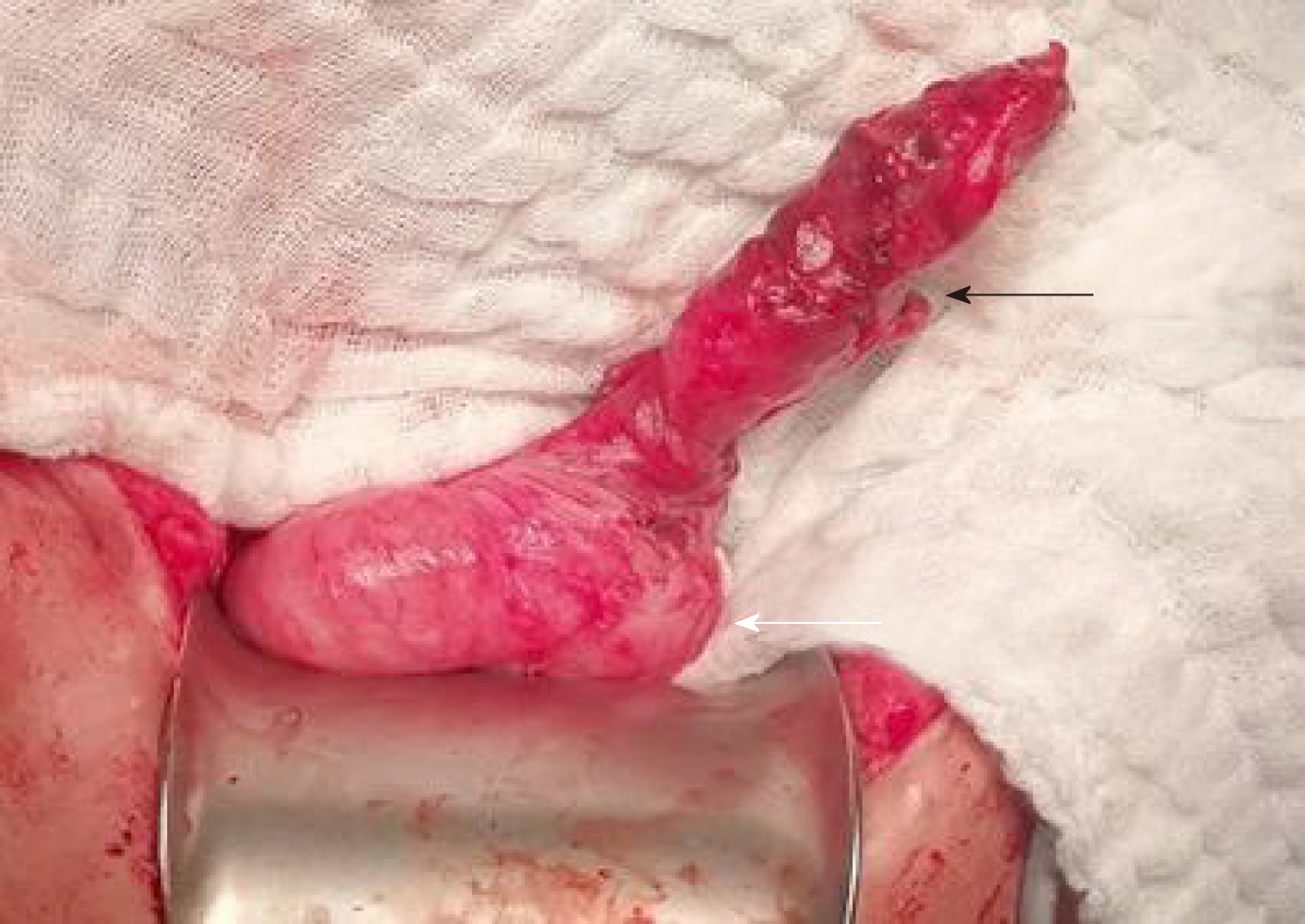
Figure 3 A photo of the appendix.
A perforation of the appendix (black arrow); the base of the caecum (white arrow).
Figure 4 A histological section of the appendix; acute gangrenous appendicitis with perforation of its wall, purulent fibrinous periappendicitis.
(Hematoxylin eosin, magnitude 20 ×). A perforation of the appendix (arrow); the lumen of the appendix (star).
- Citation: Kristek J, Kudla M, Chlupac J, Novotny R, Mirejovsky T, Janousek L, Fronek J. Acute appendicitis in a patient after a uterus transplant: A case report. World J Clin Cases 2019; 7(24): 4270-4276
- URL: https://www.wjgnet.com/2307-8960/full/v7/i24/4270.htm
- DOI: https://dx.doi.org/10.12998/wjcc.v7.i24.4270


