Copyright
©The Author(s) 2019.
World J Clin Cases. Nov 26, 2019; 7(22): 3881-3886
Published online Nov 26, 2019. doi: 10.12998/wjcc.v7.i22.3881
Published online Nov 26, 2019. doi: 10.12998/wjcc.v7.i22.3881
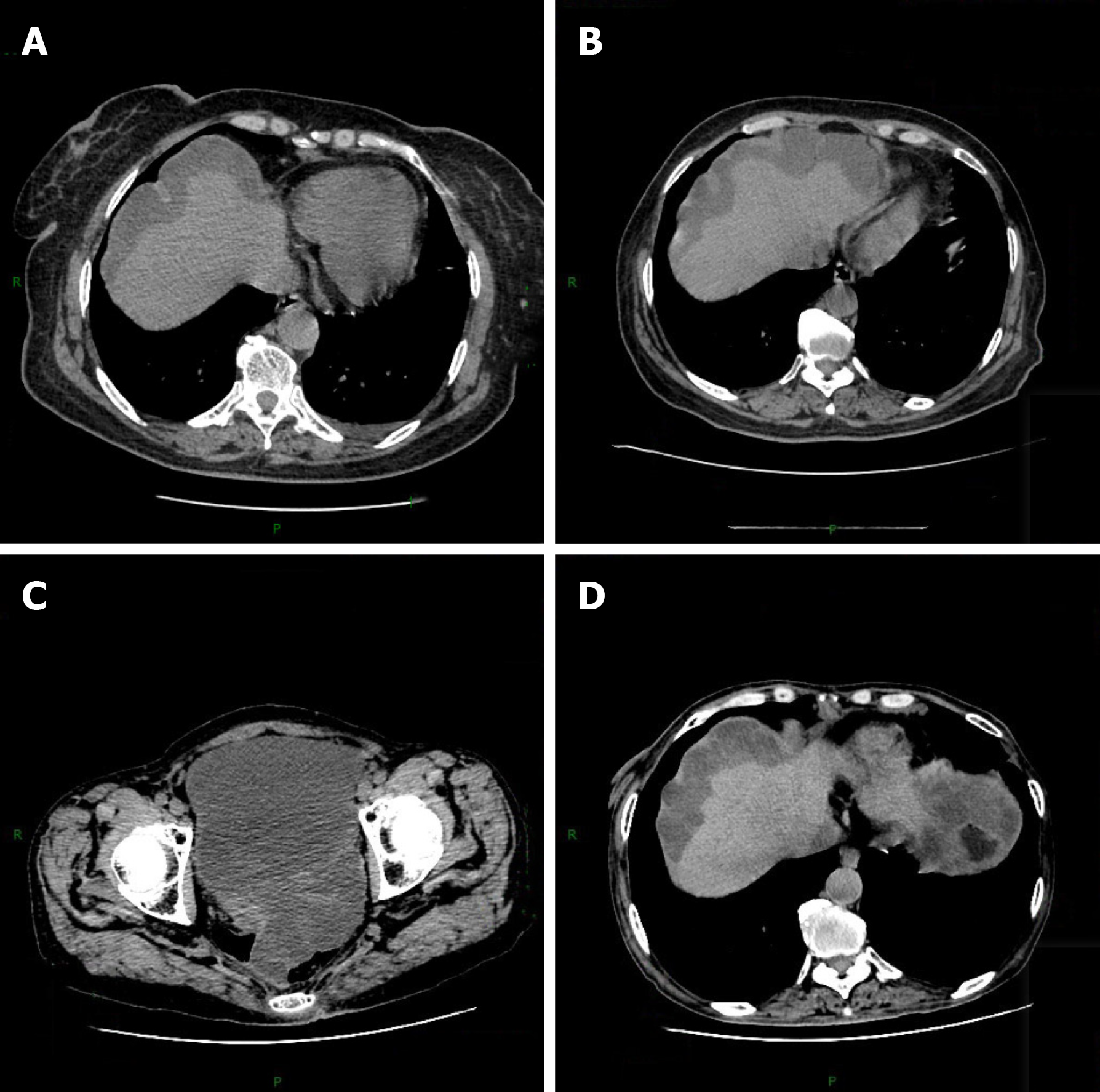
Figure 1 Chest and abdomen computed tomography.
A: Before the operative treatment (November 24, 2015); B: After the operative treatment (January 1, 2016); C, D: After the apatinib treatment (December 18, 2017).
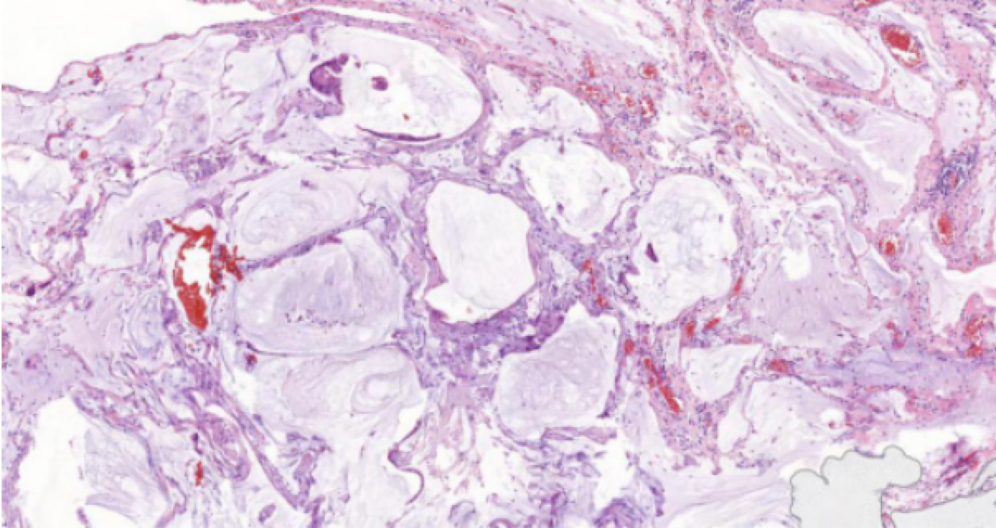
Figure 2 Hematoxylin and eosin staining of appendix lesions (×200).
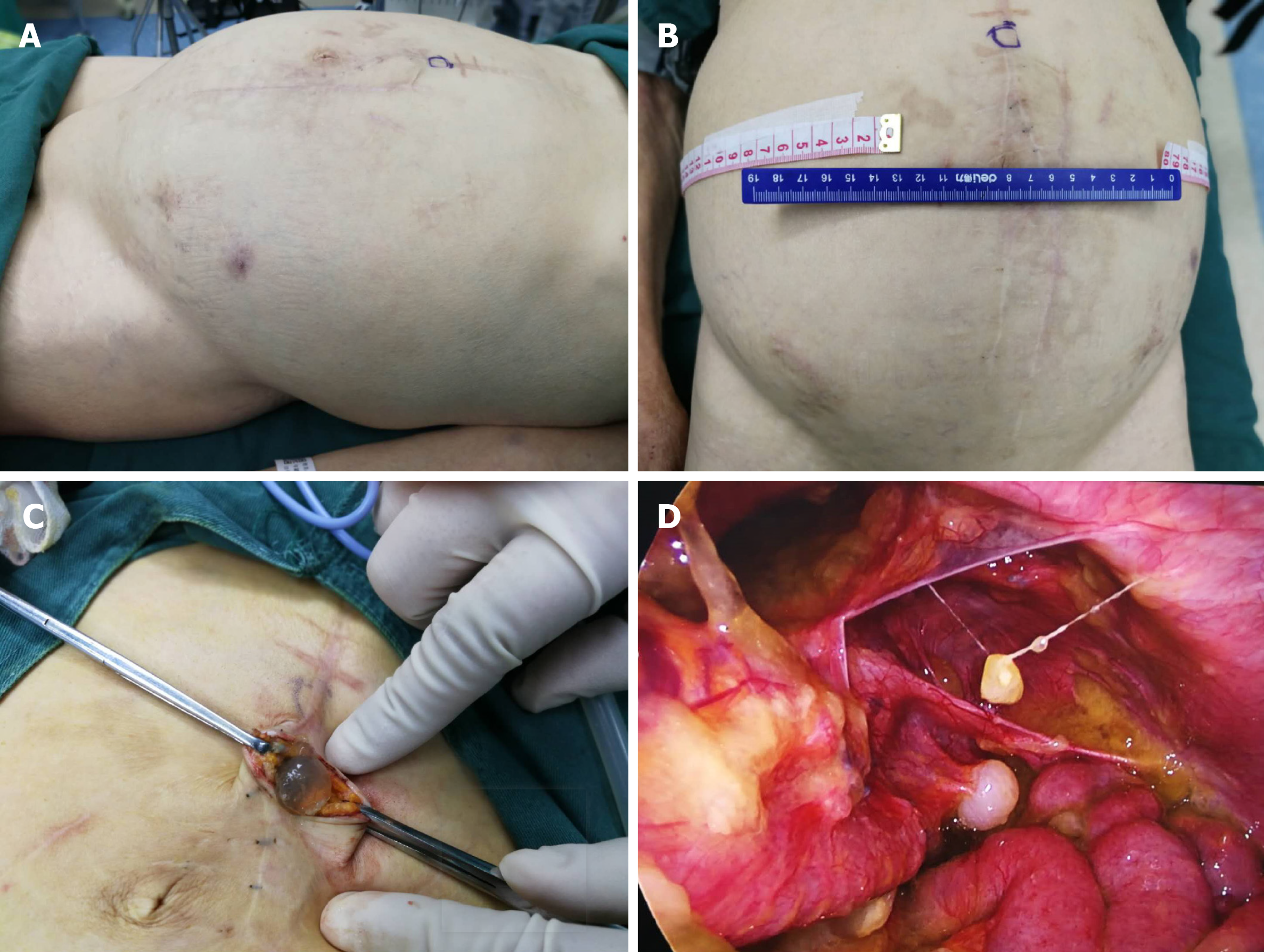
Figure 3 Preoperative and intraoperative photos of the patient.
A: Preoperative side; B: Preoperative front; C: Cutting skin; D: Entering the abdominal cavity.
- Citation: Huang R, Shi XL, Wang YF, Yang F, Wang TT, Peng CX. Apatinib for treatment of a pseudomyxoma peritonei patient after surgical treatment and hyperthermic intraperitoneal chemotherapy: A case report. World J Clin Cases 2019; 7(22): 3881-3886
- URL: https://www.wjgnet.com/2307-8960/full/v7/i22/3881.htm
- DOI: https://dx.doi.org/10.12998/wjcc.v7.i22.3881


